What are stabilizers?
There are 2 muscle groups in the human body: motors and stabilizers. The former are responsible for the mobility of individual parts of the body.
Stabilizers are a group of muscles that fix the body in a certain position (they do not take part in movement) and protect against various types of damage.
This category includes:
- abdominal muscles (transverse, rectus);
- gluteal (small, medium);
- adductor muscles;
- subosseous;
- thigh muscles (back surface);
- coracobrachial.
Stabilizing muscles are located in fairly deep layers of the human body. They are small in size. However, this does not diminish their importance. The functions they perform are so important, especially for athletes, that there is no doubt about the need to train them.
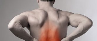
Causes of damage
The knee ligaments are very powerful structures. To damage them, you need to apply significant force. However, despite all the strength of these connective tissue structures, their injury, unfortunately, occurs quite often.
The nature of damage to the lateral and knee joints can be different - from stretching or tearing of individual fibers to their separation from the place of attachment, sometimes even with a bone area. Rupture of these fibrous structures can often be triggered by injuries and excessive stress on the joint, which occurs most often in professional athletes.
The cause of rupture or even separation of the internal collateral ligament from its attachment site can also be enthesopathy - damage to fibrous structures that is both inflammatory and degenerative in nature. It does not occur on its own, but is a consequence of destructive changes in the joints - spondyloarthritis, psoriasis, rheumatoid arthritis.
Quite often, the lateral (collateral) ligaments are injured when there is a sharp forceful deviation towards the straightened shin. Injury to the knee joint most often occurs with lateral deviation of the tibia and a bent knee with simultaneous rotation outward or inward.
Do they need to be developed?
Of course, a beginner will have a completely natural question: “Why do you need to train the stabilizer muscles?”
If we talk about sports, these fabrics perform many necessary functions. For example:
- A ski racer uses stabilizers to maintain balance under the influence of a wide variety of factors.
- These same muscles help you lift more weight during strength training. When doing a standing press, no matter how developed the triceps and shoulders are, the load mainly falls on the tissues of the lower back, which provide support for the body.
These muscles are no less important in everyday life:
- Walking and climbing stairs require maintaining balance. To do this, you simply need a fixing ability.
- A sedentary lifestyle leads to severe back pain. The cervical region suffers, discomfort is felt in the thoracic region, and the lower back makes itself felt. Doctors say that such pain is dictated by the underdevelopment of stabilizers, which are not able to properly support the spine.
- Most people dream of excellent muscle definition. This issue is especially interesting for women. To provide the body with relief, it is not enough to exercise only the superficial muscles. It should not be forgotten that the frame is formed by stabilizer muscles. That is why it is necessary to take care of training for them.
Now let's look at the most effective exercises that can strengthen these muscle groups.
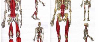
Knee joint: anatomy and physiology of the muscular system
Alternate contraction and relaxation of the muscles causes the knee to move in three planes, thereby ensuring mobility and stability of the lower limb. That is why the main classification of the muscular system is based not on the anatomy or localization of each group, but on the functions assigned to it:
- Knee flexion. This movement is ensured by the balanced and complete work of the largest muscle group of the knee joint. It includes the biceps, semitendinosus, semimembranosus, popliteal, gastrocnemius, plantaris, sartorius and gracilis muscles.
- Extension of the joint. This function is assigned to only one, but the largest muscle of the leg - the quadriceps. It consists of the direct, lateral, medial and intermediate wide muscle fibers.
- Pronation is the inward movement of the leg. Limited “rolling” of the lower leg towards the internal axis is ensured by the popliteal, semitendinosus, gracilis, sartorius, semimembranosus, and medial head of the gastrocnemius muscles.
- Supination is an outward movement. Turning the tibia outward is possible due to the contraction of the biceps and lateral head of the gastrocnemius muscle.
Innervation of tissues adjacent to the knee joint
The nerve fibers of the knee joint are a complex interconnected network, which ensures the full functioning of the lower extremities. Despite the fact that the innervation network of the knee is not very developed, each of its elements plays a key role, which means that at the slightest failure the entire system of joint mobility is “switched off”.
The nervous system, localized in the knee area, is represented by the following fibers:
- The meniscal nerve bundles penetrate the tissue along the periphery of the body of the cartilage itself, along the blood vessels of the knee. These nerves contribute to the formation of soft and soft fibers, maintaining normal innervation of joint tissues.
- The tibial nerve, through its articular branches, provides sensation to the posterior surface of the knee.
- The peroneal nerve innervates the anterior part of the knee, including the cap.
Anatomy of the blood vessels of the knee
The two key blood vessels located in the area of the knee joint are localized on the posterior surface, that is, under the knee (which is why both the vein and the artery are called popliteal in anatomical reference books). The artery transiently carries blood from the heart to the lower parts of the leg - the leg and foot, and the vein of the same name, in turn, returns depleted blood to the heart. However, these vessels do not represent the entire circulatory system of the knee: many vessels of smaller diameter depart from them, connected to each other by a network of anastomoses. Thanks to them, nutrition is provided to the muscles and tissues adjacent to the knee joint.
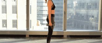
Exercise "Plank"
Take a position as if you were preparing to do push-ups on the floor. Arms should be straight. Don't arch your back. You need to stay in this position for a few minutes. You should feel the inner muscles tense. This is the basic level. It perfectly trains the spinal stabilizer muscles.
Exercises based on the “Planck” can vary and become more complex as the athlete prepares:
- plank with emphasis on elbows;
- emphasis on the elbows and raising one leg;
- side stand;
- support on one arm/leg.
Treatment of injuries
An integrated approach is usually used to treat such conditions. Enthesopathies, as well as ruptures and sprains of connective tissue structures, need to be treated using various approaches:
Enthesopathy of the medial collateral ligament is a condition that cannot be treated. The disease that caused it needs to be treated.
Knee ligament rupture
The rupture may be complete or partial. Depending on this, it can be treated conservatively or surgically.
Conservative treatment
This treatment includes puncture of the knee joint, after which a plaster cast is applied for 1.5 months in the position of the greatest possible deviation towards the affected fibrous structure. This treatment is effective for tears, for example, of the medial collateral ligament.
However, when it is completely ruptured, this structure does not always heal, and the external collateral ligament does not heal at all. When it ruptures, surgical treatment is definitely necessary. It makes no sense to treat old ruptures (occurring more than 2-3 weeks ago) of the medial collateral ligament conservatively.
If the knee joint is damaged, sometimes, depending on how much the instability of the knee joint is compensated, it is possible to prescribe conservative treatment (application of a plaster splint or orthosis for up to 6 weeks).
In order to reduce swelling and relieve pain, the doctor prescribes an appropriate ointment or compress.
Surgical treatment
Even with complete ruptures of the anterior knee joint, patients who lead a sedentary lifestyle are satisfied with the results of conservative treatment. If a person leads an active lifestyle, then with a complete rupture of the joint, only surgical treatment is indicated.
Operations can be performed using several methods:
- Arthroscopy.
- Arthrotomy (when the knee joint is opened and the necessary manipulations are performed under visual control).
Surgery for a complete rupture of the lateral collateral ligament should be carried out in the first three days after the injury and consists of stitching the separated fibers of the damaged structure together. If time has been lost, depending on the situation, if the collateral ligaments are damaged, plastic surgery is performed using a section of one’s own tissue or with a special polymer linen.
After the operation, it is necessary to apply a fixator, for example, a plaster splint (a splint from the fingers to the upper third of the thigh) or a hinged orthosis for 1.5 months. 21 days after application, and after consulting with your doctor, it is advisable to begin working out the knee joint to prevent the development of stiffness in it.
Surgery for a ruptured knee joint is performed in the first three days after the injury or, which happens much more often, several weeks after the injury.
Depending on the duration of the injury, fragments of this structure will either be sewn together, or their plasticity will be performed using one’s own tissues or using artificial materials.
After surgery, a hinged orthosis or cast is applied to almost the entire leg for up to 6 weeks.
Knee sprain
How to cure a knee sprain? Surgery is not necessary to treat a knee sprain, but arthroscopy may be necessary for hemarthrosis. Such injuries should be treated based on the following principles:
- Rest - it is necessary to unload the affected limb as much as possible. To do this, you don’t need to try to stand on it or bend it.
- Cold – in the first day after injury, it is necessary to apply ice to the affected joint. Ice wrapped in cloth is applied for 10–15 minutes, then removed.
- Fixation - the joint is fixed with an elastic bandage or another non-rigid fixator, unless the doctor prescribes anything else.
- Elevated position of the affected limb - to reduce swelling, it is recommended to keep the leg elevated (for example, using a pillow).
Starting from the second day of the disease, if there are no other doctor’s prescriptions, painkillers, ointments and compresses are used. The ointment recommended in these cases can be both anesthetic and warming.
Complex "Push-ups"
The exercise is performed in a variety of variations:
- Hand placement. During push-ups, you can spread your upper limbs wide apart. The muscles will be pumped no less effectively if the hands are placed side by side (“grasshopper”).
- Positioning the legs. Perform the exercise with your lower limbs wide apart on a support. Now use a narrow foot stance. Rest your lower limbs on the bench. Try lifting one leg while doing push-ups.
- Unstable support. Such exercises are recommended for professionals. You can lean your hands or feet on the rope.
You should feel how various stabilizer muscles work: back, abdomen, hips.
Exercise "Squat"
There are also various variations for this activity:
- "Pistol." Do squats on one leg. To make it more difficult, you can place the second limb on a stable support or an unstable one. Use TRX loops or their equivalents.
- Regular squats. The exercise is performed on two legs. However, use a narrow or unstable support. You can squat on a bosu (hemisphere).
Complex “Gluteal bridge”
This is a great exercise that will pump up the stabilizer muscles of the spine, buttocks, and abdomen.
The basic version works like this. Lie on your back. Hands - palms to the floor, along the body. Legs are bent at the knees, feet pressed to the floor. Without lifting your head and shoulders, lift your pelvis. Stay in this position for a few minutes.
Advanced athletes can make the exercise more difficult:
- support on only one leg;
- use a hill (bench, unstable support) for the lower extremities.
Exercise "Wheelbarrow"
This complex involves walking exclusively on your hands (your legs must be supported by an assistant). In this case, it is necessary to move forward and then backward. To make this activity more difficult, an assistant can support the athlete with only one leg. This makes it very difficult to hold the body in the required position. But at the same time, the load on the stabilizer muscles is simply colossal.
It has been noticed that the “Wheelbarrow” exercise always increases the athlete’s emotional background. In addition, this complex can be performed for a while, presented as a relay race.
No crunches needed?
Research shows that exercises such as crunches, which pull the lumbar spine into flexion, place a lot of pressure on the spine. However, I have yet to see anyone strain their back with crunches, so if you enjoy them they are probably unlikely to cause injury. In fact, crunches are good for bodybuilders who want to maximize rectus abdominis hypertrophy.
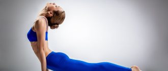
A complex that trains the back stabilizer muscles
Exercises to strengthen the corset are performed on the floor, lying on your stomach.
The complex includes:
- Alternately (simultaneously) lift your legs and head and chest off the floor surface. Perform the exercise without delay in the upper position. Then repeat, including a small stop at the top.
- Lift your right arm and left leg off the floor. Be sure to stay late. Then lower yourself to the floor. Repeat the exercise for your left arm and right leg.
- Lying on the floor, imitate the swimming technique.
The best workouts to develop stabilizer muscles
Above was a complex that does not require sports equipment. However, experts have developed certain simulators that allow you to improve exercises for the stabilizer muscles.
The most effective:
- Bosu. This is a special hemisphere simulator. It has an elastic “dome”. Exercises are performed on it in a sitting, lying, standing position (one/two legs). This platform is quite unstable. To maintain balance while on it, you need to tense almost all the muscle tissue of your core.
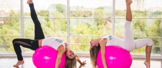
- Fitball. This is a popular accessory for sports. It is a big elastic ball. You can sit on it, lie down or lean on it. At the same time, it will constantly roll back. Use this exercise machine for push-ups (it will become a support for your legs) and abdominal pumping.
- Medball. It is also called a medicine ball. The dimensions of a medicine ball are much smaller than a fitball. It resembles a basketball. But it is distinguished by its noticeable weight - from 1 kg to 27. They use a medicine ball for push-ups and abdominal crunches.
- TRX hinges. An excellent product that strengthens stabilizer muscles. This versatile exercise machine develops endurance, strength, improves coordination, and builds balance and balance. Using this simulator, you can complicate a variety of exercises.
Exercises with sports equipment
Access to quality equipment for developing stabilizing muscles is quite easy. You don’t even need access to a fitness club - you can easily buy all the equipment listed below at any sports store and start training at home today.
Examples of exercises on a fitball
— rest your knees on the floor, fitball in front of you, hands on top of the ball; moving the body forward, the projectile is pushed forward, the angle between the body and the floor decreases, the core muscles are tense, the body is in one line from the knees to the shoulders
- triceps push-ups: from the reverse plank position, the arms are bent at the elbow joints, the body goes down, rises to the starting position
— in the boat pose, the fitball is clamped between the feet, the legs are raised up to a right angle, the body is twisted, the shoulders are lifted higher from the floor, the ball is passed from the feet to the palms, the body returns to its original position, the exercise is repeated, the ball is passed from the hand to the feet.
Examples of bosu exercises
Bosu - is a hemisphere with an elastic dome, as follows from the abbreviation (both side use) can be used by both sides, the device creates significant instability, stimulating the work of a large number of stabilizer muscles.
On the Bosu, installed with the hemisphere up, basic exercises are done: squats, leg swings, boat, planks, gluteal bridge; on the installed hemisphere down - push-ups.
- standing on the platform with emphasis on the knees and palms, the opposite arm and leg are abducted alternately
- while supporting the hands on the platform with the hemisphere down, jumping jacks are performed from the plank position
— performing a V-fold, statically or dynamically
- static chair exercise on Bosu
Unlike a fitball, Bosu is more stable, which means the risk of falling and injury is much lower, and provides a significantly larger number of exercise variations, because You can stand on it with your feet, and not just lean on it while sitting or lying down.
There are some restrictions on working with the platform: level of training - many exercises are not designed for beginners, working with weights - if you lose balance you can hit yourself with dumbbells, so the exercises are performed with your own weight.
Examples of exercises on TRX (suspension loops)
TRX loops are sports equipment that uses all muscle groups, with the help of which the body works in different planes; unlike conventional exercise equipment, it does not provide support in the form of a seat or backrest. It is impossible to perform exercises with TRX without engaging the stabilizer muscles.
With TRX it is possible to perform all the usual basic exercises, while the support of the arms or legs occurs not on a stationary surface, but on movable hinges
— reverse pull-ups: the classic exercise is performed with a loop grip
Reverse Pull-Ups
- pistol squat with loop grip
- lifting the pelvis up from the plank position
Board balance exercises
Came from training for sports such as snowboarding and skateboarding. The search for an equilibrium position on the apparatus involves all muscle groups responsible for stabilization and develops the vestibular apparatus.
Reviews from people
You can develop stabilizer muscles at any age. Such training is suitable for children, they will be an excellent support for the adult population, and will bring invaluable benefits to the elderly. People testify that the development of stabilizer muscles can significantly strengthen the back corset. And this, in turn, relieves the population from unpleasant back pain.
However, despite the obvious benefits, these complexes have some contraindications. Therefore, people who have problems with joints or spine should definitely consult with a competent doctor.
Development of stabilizer muscles[edit | edit code]
Source: "Training Programs"
, scientific ed.
Author:
Professor, Doctor of Science Tudor Bompa, 2020
Propulsion muscles work more effectively when combined with strong stabilizer or anchor muscles. Stabilizer muscles contract primarily isometrically to stabilize a joint so that an action can be performed by another part of the body. For example, during elbow flexion, the shoulders are fixed in a stationary position, and the abdominal muscles act as stabilizers when throwing a ball with the hand. When rowing, when the muscles of the torso act as stabilizers, the torso transfers the power of the legs to the arms as the oar passes through the water. Accordingly, weak stabilizer muscles block the ability of driving muscles to contract.
If the stabilizing muscles are not properly developed, the core muscles may also become impaired. With chronic exercise, spasm of the stabilizer muscles may occur, which limits the work of the moving muscles and reduces the athlete’s performance. A similar situation is often observed in volleyball players who are injured as a result of poor muscle strength and imbalance in the muscles of the shoulder girdle[1]. The infraspinatus and supraspinatus muscles of the shoulder girdle are responsible for rotating the arm. The simplest and most effective way to strengthen these muscles is to rotate your arms with dumbbells. As a result of the resulting resistance, two muscle groups that stabilize the shoulder are stimulated. External (external) rotation of the hip joint is carried out due to the work of the piriformis and gluteus medius muscles. To strengthen these muscles, the athlete must take a standing position with fixed knees and lift the leg to the side using the load from the crossover exercise machine.
Stabilizing muscles also contract isometrically, resulting in immobilization of one part of the limb and movement of the other. In addition, these muscles control the interaction of the long bones in the joints and signal the risk of injury as a result of improper exercise technique, improper strength, or spasms that occur due to lack of stress management. If one of these conditions is met, the stabilizer muscles limit the work of the prime mover muscles, preventing the risk of strain or injury.
The above suggests that stabilizer muscles play an important role in improving the performance of athletes. On the other hand, some strength and conditioning coaches have recently placed too much emphasis on developing stabilizer muscles, especially through proprioception training (also called balance training). In fact, training on an unstable surface causes activation of the upper motor units as a result of simultaneous muscle contraction (meaning the simultaneous contraction of agonist and antagonist muscles to stabilize the joint), which does not lead to the neuromuscular adaptations that are necessary for athletes participating in speed-strength sports. These athletes require that antagonist muscles (i.e., passive muscles) be at rest during forceful activities.
On the other hand, many studies have shown that proprioception training using balance boards helps strengthen unstable or previously injured ankles[2][3][4]. Current theory suggests that if balance board training improves stability by improving proprioception and strengthening the stabilizer muscles of an unstable structure, the training will also provide further muscle strengthening and reduce the risk of muscle injury by providing a stable structure. However, this effect has not yet been proven, and the question of how much time should be devoted to exercises designed to strengthen stabilizer muscles remains unresolved.
Some studies suggest that proprioception may reduce the risk of knee injury[2], while other studies argue against the usefulness of proprioception training as a means of injury prevention[5]. In particular, a recent peer review study examined pitfalls in the design and implementation of proprioceptive learning outcomes[6]. Additionally, reports from strength and conditioning coaches who completely ignored the use of balance boards or proprioceptive training in team sports (soccer and volleyball) suggest that knee and ankle injuries remained unchanged in over the past 10 years.
Taking into account the above, we can conclude that training using a gymnastic ball or balance board can be useful in the early stage of the preparatory stage (anatomical adaptation stage). Of course, unilateral exercises are the optimal choice for improving joint strength while training the prime mover muscles. Proprioception training is good for the anatomical adaptation stage, but at a later stage the exercise ball and balance board should be put aside to free up time to train the athlete using techniques that directly improve his physical condition and promote the development of specific strength, speed and endurance. In general, even if the athlete’s proprioception has improved as a result of the exercises, the regularity and slowness of these exercises do not protect the joint from sudden and powerful movements performed during sports [7]. Preparing the stabilizing muscles for movement plays an important role: in particular, to ensure high performance and optimal fitness of the athlete, it is vital to train the movements specific to a particular sport at the appropriate level of specific speed, strength and endurance.
The table shows the breakdown of a young football player's work schedule over three weeks during the macrocycle of anatomical adaptation. Pay attention to the large number of unilateral exercises, the same amount of work performed by the agonist and antagonist muscles, the duration of the sets under load, which does not exceed the range of the lactate system (from 48 to 80 seconds), gradually increasing the load and shorter duration macrocycle, which is typical for young athletes and professionals. Below is a point-by-point description for each column in the figure:
- sets: each number represents the number of sets performed during a particular week. For example, 2-3-2 means two sets are performed during the first week, three sets during the second week, and two sets during the third week;
- Repetitions: Each number represents the number of repetitions performed during a particular week. For example, 20-15-12 means 20 reps per set are performed in the first week, 15 reps per set in the second week, and 12 reps per set in the third week;
- Rest Break: Each number represents the length of rest between sets of exercises for a given week. For example, 1-1-1.5 means that during the first week the rest break between sets lasts one minute, during the second week - one minute, during the third week - one and a half minutes.
- repetition time: the first digit displays the duration of the eccentric phase in seconds, the second digit displays the pause between the eccentric and concentric phases, and the third digit displays the duration of the concentric phase (X indicates the explosive phase);
- Load: These columns are used to record the weekly load for each set of each exercise.
Rough breakdown of a young football player's three-week work schedule during the macrocycle of anatomical adaptation
| Exercise | Approaches | Repetitions | Rest break (min.) | lead time | LOAD | ||
| 1 Week | 2 week | 3 week | |||||
| Workout A | |||||||
| Single leg squats | 2-3-2* | 20-15-12* | 1-1-1,5* | 3-0-1** | |||
| Single leg curl | 2-3-2 | 20-15-12 | 1-1-1,5 | 3-0-X | |||
| Single leg deadlift | 2-3-2 | 20-15-12 | 1-1-1,5 | 3-0-1 | |||
| Extension of the gluteal muscles and quadriceps | 2-3-2 | 20-15-12 | 1-1-1,5 | 3-0-1 | |||
| Working with abductor muscles on the simulator | 2-3-2 | 20-15-12 | 1-1-1,5 | 3-0-1 | |||
| Working with adductor muscles on the simulator | 2-3-2 | 20-15-12 | 1-1-1,5 | 3-0-1 | |||
| Standing calf raise | 2-3-2 | 20-15-12 | 1-1-1,5 | 2-2-1 | |||
| Crunches using weights | 2-3-2 | 20-15-12 | 1 | 3-0-3 | |||
| Workout B | |||||||
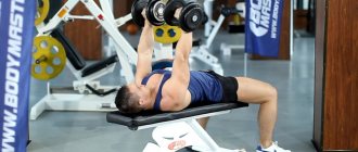
| Dumbbell supine press | 2-3-2 | 20-15-12 | 1-1-1,5 | 3-0-1 | |||
| Dumbbell row | 2-3-2 | 20-15-12 | 1-1-1,5 | 3-0-1 | |||
| Seated dumbbell press | 2-3-2 | 20-15-12 | 1-1-1,5 | 3-0-1 | |||
| Dumbbell biceps curl | 2-3-2 | 20-15-12 | 1-1-1,5 | 3-0-1 | |||
| Front and side elbow stand | 2-2-1 | 30-30—45 (sec.) | 0,5 | Isometric | |||
*For each triple of digits in this column, the first digit refers to the first week, the second digit refers to the second week, and the third digit refers to the third week.
**In each triple of numbers in this column, the first number represents the duration of the eccentric phase in seconds, the second number represents the duration of the pause between the eccentric and concentric phases, and the third represents the duration of the concentric phase (X indicates the explosive phase).