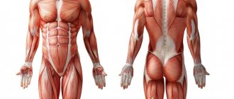
Muscle hyperplasia[edit | edit code]
The difference between hypertrophy and hyperplasia
Hyperplasia
(Novolat. hyperplasia; other Greek. ὑπερ- - super- + πλάσις - formation, formation) - an increase in the number of structural elements of muscle tissue (muscle fibers) by dividing them. Unlike hyperplasia, hypertrophy involves an increase in the volume of cells and sarcoplasmic structures, without pronounced division (new formation of nuclei).
Research[1] confirms that the contribution of hyperplasia to muscle volume is less than 5% and is more significant only when using anabolic steroids. Myostatin blockers can also cause hyperplasia. Growth hormone does not cause hyperplasia.
People prone to hypertrophy usually have a larger number of muscle fibers. The total number of fibers is determined genetically and practically does not change throughout life without the use of special pharmacology. There has been no evidence of an increase in the number of muscle fibers (muscle fiber hyperplasia) in humans under the influence of strength training, although muscle fiber hyperplasia is possible in animals (mammals and birds)[2].
Research[edit | edit code]
While during muscle hypertrophy an increase in the number of nuclei in a muscle cell is observed, in animal studies of atrophy processes the opposite phenomenon was noted. A decrease in the number of nuclei occurs as a result of atrophy of muscle fibers after transection of the spinal cord [3], during prolonged stay in conditions of weightlessness [4] or temporary immobilization of the hind limb [5]. Thus, changes in the number of nuclei per muscle cell appear to be of great importance in regulating cellular fibril size. At the same time, it must be borne in mind that an increase in the number of nuclei in a muscle fiber will occur as long as the activity of converting existing nuclei is capable of ensuring an increase in their number. Indeed, noticeable changes in the number of nuclei in the muscle fiber were observed in muscles hypertrophied by more than 26%[6], but not in muscles hypertrophied by 6.8-15.5%[7].
Since differentiated muscle fiber nuclei are unable to divide, the main source of new nuclei in hypertrophied muscle fibers are myosatellite cells or satellite cells[8]. Myosatellitocytes are located between the basal plate and the plasma membrane of muscle fibers[9], they are characterized by a high nuclear-cytoplasmic ratio, a well-developed Golgi apparatus, a pronounced granular endoplasmic reticulum and a heterochromatinized nucleus[10]. Activation of satellite cells can occur under the influence of a number of stimuli, after which they begin to actively divide. After this, the daughter cells formed as a result of mitosis merge with nearby differentiated muscle cells, thus providing an increase in the number of nuclei. The role of the nuclei formed during the division of myosatellite cells in the process of muscle hypertrophy is also confirmed by experiments on animal models demonstrating that activation and subsequent proliferation of satellite cells are necessary to ensure muscle growth [11].
It has been shown that, in parallel with muscle hypertrophy, intense strength training induces a significant increase in the number of satellite cells in skeletal muscles[12]. A 46% increase in the proportion of myosatellite cells in the skeletal muscle of a young woman was reported after 10 weeks of resistance training[13]. Recently, an increase in the number of myosatellite cells was found in the skeletal muscles of a group of men aged 70–80 years who were engaged in endurance training[14]. Thus, satellite cells ensure an increase in the number of nuclei in the muscle fiber and the renewal of their own pool [15]. Newly formed muscle fibers replace damaged ones or contribute to muscle fiber hyperplasia only if the number of newly formed fibers exceeds the number of fibers damaged during exercise.
Causes and types
What causes muscle wasting? The main cause of muscle wasting is insufficient supply of nutrients. A decrease in muscle mass can occur as a result of infectious diseases, exposure to toxins from burns, frostbite, chemical poisoning, and intoxication due to long-term compartment syndrome. Hypotrophy of the leg muscles in an adult develops when peripheral nerves or tendons are damaged. Muscle wasting occurs with paralysis.
There are several types of muscle wasting. Congenital hypotrophy of the leg muscles occurs due to pregnancy pathology (impaired blood supply to the fetus, infectious diseases of the mother, poor nutrition and bad habits of a pregnant woman). The acquired form of the disease develops after birth trauma, with unbalanced nutrition of the child, metabolic disorders, and dysfunction of the endocrine organs. Muscle wasting of the lower extremities in old age occurs when a person is insufficiently active.
The most common type is hip muscle wasting in adults. It develops when the function of the hip joint is impaired and coxarthrosis develops. Lower leg hypotrophy most often occurs in young children who do not receive a balanced diet and are often injured. Athletes who have stopped exercising are at risk of developing muscle wasting. Leg hypotrophy develops in office workers, cashiers, and people who spend most of the day at the computer. Muscle wasting is characteristic of damage to the peripheral nervous system.
Muscle wasting accompanies flaccid paralysis, which occurs with the paralytic form of poliomyelitis. Muscle atrophy develops gradually with: the following pathology:
- Hereditary degenerative diseases of the muscular system;
- Metabolic disorders;
- Chronic infections;
- Long-term use of glucocorticoids;
- Violations of the trophic functions of the nervous system.
Local muscle wasting occurs with prolonged immobility associated with a violation of the integrity of tendons, nerves or muscles, or joint diseases.
Hypertrophy or hyperplasia?[edit | edit code]
Source
: Iron World. No. 6.2013
What is the reason for the growth of muscle mass, hypertrophy of muscle fibers (an increase in the volume of muscle fibers), or their hyperplasia (an increase in the number of muscle fibers)? Official science does not confirm the data on the possibility of CF hyperplasia in humans, although there are quite a lot of facts confirming the presence of this process in animals. In recent years, however, works have begun to be published frequently that question the official point of view.
Supporters of CF hyperplasia were supported by such a well-known and deservedly respected specialist in power sports as Mikhail Klestov, well known to readers of our magazine:
- The limit to the possibility of hypertrophy
is doubling the diameter of the muscle fiber. The most massive athletes have muscle fibers no more than 2 times thicker than those of the leanest dystrophic person. Further progress is possible only due to hyperplasia. The muscle spindle can increase its diameter by a maximum of two times. This is due, among other things, to problems of a trophic nature. At least, science does not know a single fact that a muscle spindle is three times larger than the average diameter. A maximum twofold increase in diameter was observed. However, among the pros there are many athletes who have increased their muscle mass three times or more.”
Very logical, isn't it? But only if you don’t doubt the original data. Unfortunately, I have too often witnessed the extreme inertia reigning in science. For example, an important discovery has been made in the field of physiology, which allows us to completely rethink and change the existing stereotype in training. Do you think everyone will start making adjustments right away? No. Firstly, in order for people to know about this information, they need to invest huge amounts of money in its promotion. Scientists themselves do not do this; their job is scientific work. If a brilliant discovery does not immediately bring huge dividends, then there are not many people willing to spread information about it or somehow promote it. Moreover, it is often met with hostility. About fifteen years ago, at the Research Institute of Fundamental and Applied Problems of Physical Education and Sports, the possibility of local fat burning was irrefutably proven. Proven, scientifically substantiated and confirmed by a huge amount of statistical material. All this was published in scientific publications, but the inertia of science is such that even now at training courses for fitness trainers and seminars they continue to repeat the impossibility of local fat burning. The indications of such types of testing as body mass index (BMI) and the Karvonen test have long been considered unscientific and false, but, nevertheless, they are stubbornly implanted in all fitness centers.
Unfortunately, in our country we do not have a service that would monitor all serious scientific discoveries published in scientific journals and introduce our specialists to these discoveries. Therefore, we decided to turn to Professor Viktor Nikolaevich Seluyanov for clarification on this issue, who has been studying and analyzing all the major scientific periodicals in the world for several decades.
Iron World
: Viktor Nikolaevich, what is the main cause of muscle growth, hypertrophy or hyperplasia of muscle fibers? Has there been any research done in the world that remains unknown to our coaches and strength training specialists?
Victor Seluyanov
: In the 70-80s, the question arose of what causes muscle growth in athletes, especially bodybuilders. Then they took biopsies from athletes, and indeed it turned out that the cross-sectional size of the muscle fiber in bodybuilders was only 30% larger than in ordinary people. And the appearance of an ordinary person and a bodybuilder differs significantly. 3, and maybe 4 times. Therefore, they began to look for the reasons why such muscle growth is possible. In the USSR you can find such an author as Druzd, who began to study the muscles of trained people using biopsies and in the end he found that large muscle fibers can divide. The so-called longitudinal division occurs in them, which is possible; in addition to increasing the size of the muscle fiber, some laws arise according to which their bifurcation occurs. Thus, the number of muscle fibers increases. At the same time, there was no talk about myosatellites. By the way, myosatellites still belong to an unknown area of knowledge. It is still believed that myosatellites do not participate in CF hypertrophy, that is, new fibers are not formed due to myosatellites. Although now they are trying to influence them with the help of pharmacology and thus increase muscle volume, we will assume that this is still a hypothetical area. Time has passed. More and more biopsy samples were taken from athletes of varying fitness levels. If previously it was believed that only 30% of the fibers could be hypertrophied, then subsequent studies showed that the size of the muscle fiber can be increased by 3-4-6 times or more! And the factor of the possibility of CF hyperplasia moved aside. Today we can clearly say that the number of muscle fibers in a person is set from birth. If one person grows muscles quickly, it is not because the amount of MV increases in him, but he initially had a lot of MV, and another, even if he wants to, will not be able to build up large muscle mass, because he initially has little MV.
ZhM
: But if he can increase their diameter by 6 times.
Victor Seluyanov
: Yes, it can increase, but it cannot reach a high level in bodybuilding. He will still lose to an opponent who has 3 times more muscle fibers. In speed skating, for example, the prospects of an athlete are determined by the size of the quadriceps femoris muscle. If this muscle is not large from birth, then he will not get good results in skating. And if it is initially large, then in a year or two he is able to fulfill the MS standard. There are many such cases. One of the most famous is speed skater Oleg Goncharenko. In two years of training he became a world champion. But he came with huge legs.
ZhM
: So, you completely deny the possibility of CF hyperplasia?
Victor Seluyanov
: Perhaps this factor exists. I think up to 3% muscle fiber can be added. One can agree with Druzd regarding longitudinal splitting. But no one has yet proven that CF hyperplasia can be a significant factor in increasing muscle size, increasing muscle strength, and increasing athletic performance.
ZhM
: There has been an opinion in bodybuilding for many years that CF hyperplasia is promoted by taking growth hormone.
Victor Seluyanov
: No, growth hormone, of course, does not work to increase the amount of CF. It enters cells, affects DNA, and in cells it begins to more actively build components responsible for its strength. It is especially active in tendons, ligaments, and places of attachment of muscle tissue to the tendon. Some muscle tissue also grows. Myofibril hyperplasia occurs in the muscle fiber. That's all that is currently known about the anabolic effect of growth hormone, and the rest is more fiction than scientifically explainable facts.
ZhM
: If in the late 80s - 90s studies were already carried out that proved the possibility of increasing the diameter of MVs by 6 or more times, why is there no such data in the domestic literature? And it is still stubbornly said that it is impossible to increase MV by more than 2 times
Victor Seluyanov
: Perhaps because there are not many such articles and such studies in the world. I know of 3-4 articles with similar studies. To do this, it is necessary to constantly monitor all published scientific literature in order to select 1-2 out of 1000 articles on the topic of interest, which were written at a high scientific level and on a contingent of high-class athletes. Now I will introduce you to one of the very worthy works published back in 1989. It has never been translated into Russian, my translation.
Comparative analysis of muscle fibers of elite male and female bodybuilders
[16]
The problem of the degree of hypertrophy of muscle fibers was studied on highly qualified bodybuilders. There were 8 men and 5 women. The average anthropometric indicators were 173 cm, 87 kg and 166 cm and 62 kg, respectively.
The subjects of the study were the elbow flexors, long head of the biceps brachii and brachialis. A biopsy was taken from these muscles. The tissue sample was frozen in liquid nitrogen. Muscle composition was determined according to Bergstrom. Myosin ATPase activity was assessed. The cross-section of muscle fibers was measured under a microscope (x15000). The total muscle area was measured from a photograph of a muscle section after computed tomography. The amount of MV in the muscle was determined by dividing the muscle area by the cross section of the average muscle fiber.
As a result, the share of type 2 CF (fast) was around 50%. The share of the non-contractile part was 9-10%. The average cross-sectional area was 7,200 mm2 and 4,700 mm2 for type 1, 11,400 mm2 and 5,000 mm2 for type 2 in men and women, respectively. Of particular interest are data on the cross-sectional distribution of muscle fibers. In Fig. it can be seen that the size of muscle fibers ranges from 2000mm2 to 15000mm2 in women and up to 20000mm2 in men. The shoulder girth for men was 47 cm; if we recalculate, taking into account the reduction in the cross section of the MV to the norm of an untrained person (3000-4000 mm2), then the shoulder girth will be 27-30 cm. Consequently, in bodybuilders, muscle mass growth was associated only with MF hypertrophy (myofibril hyperplasia). “There is no room left for hyperplasia of muscle fibers.”
Event frequency by area
Comments
:
The non-contractile part of the CF, that is, the non-contractile part. This is what enters the muscle cell in addition to myofibrils. This includes mitochondria, sarcoplasmic reticullum, etc. Sometimes they talk about mitochondrial and sarcoplasmic muscle hypertrophy. So they fit into this 9-10%. When professional histologists hear about these types of hypertrophies to increase muscle mass and about special training aimed at this, they start laughing. Their contribution to muscle growth can be so insignificant.
It turned out that the average cross-sectional area of elite bodybuilders in a BMW is 11,400 mm2, which exceeds the average of an untrained person (3000-4000 mm2) by 2.85-3.8 times, and in absolute values, as can be seen from the curve in the graph, The cross-sectional area of fast muscle fibers in men can exceed 20,000 mm2, which is 5 - 6.5 times higher than the average for an untrained person.
Treatment
Neurologists at the Yusupov Hospital prescribe complex treatment to patients suffering from muscle dystrophy, aimed at eliminating the cause of the disease, influencing the mechanisms of development of the pathological process, and reducing the manifestations of the disease. To improve blood flow in peripheral vessels, angioprotectors (trental, pentoxifylline, chimes), low molecular weight dextran, and prostaglandin E preparations (vasaprostan) are used. After dilating blood vessels with no-shpa and papaverine, the supply of muscle fibers with oxygen and nutrients improves.
B vitamins (thiamine hydrochloride, pyridoxine hydrotartrate, cyanocobalamin) normalize metabolic processes and the conduction of nerve impulses. Biological stimulants stimulate the regeneration of muscle fibers and restore muscle volume: aloe, actovegin, plasmol. To restore muscle conduction, prozerin, galantamine, and armin are used.
Sources[edit | edit code]
- Kraemer, William J.; Zatsiorsky, Vladimir M. (2006). Science and practice of strength training. Champaign, IL: Human Kinetics. p. 50. ISBN 0-7360-5628-9.
- MacDougall JD Hypertrophy and Hyperplasia // In: The Encyclopedia of Sport Medicine. Strength and Power in Sport. - Bodmin, Cornwall: Blackwell Publishing, 2003.
- Allen et al., 1995
- Allen et al., 1996
- Hikida et al., 1997
- Cabric, James, 1983; Allen et al., 1995; Roy et al., 1999; Kadi, Torncll, 2000
- Giddings, Gonyea, 1992
- Moss, Lcblond, 1971; Schiaffino et al., 1976
- Mauro, 1961
- Campion, 1984
- Rosenblatt, Parry, 1992; Rosenblatt et al., 1994
- Kadi, 2000; Roth et al., 2001
- Kadi, Tomell, 2000
- Charifi et al., 2003a
- Bischoff, 1994; Schultz and McCormick, 1994; Kennedy et al., 1988; Yamada et al., 1989; McCormick and Schultz, 1992; Antonio, Gonyea, 1993; Kadi, Thomell, 1999
- S. E. Alway, W. H. Grumbt, W. J. Gonyea, and J. Stray-Gundersen Contrasts in muscle and myofibers of elite male and female bodybuilders J Appl Physiol July 1, 1989 67:(1) 24-31
Diagnosis of leg muscle atrophy
Diagnosis of leg muscle atrophy is carried out by collecting a detailed history of the person, the presence of chronic diseases and hereditary burden. The patient should be referred for a complete blood test to determine the level of ESR, liver function tests, and glucose. An electromyography procedure is performed.
To determine the optimal treatment, doctors prescribe a biopsy of the nerves and muscles. At the same time, additional studies are carried out if the patient has a history of chronic or hereditary diseases.
Our specialists
Head of the Research Center for Motor Neuron Disease/ALS, Candidate of Medical Sciences, doctor of the highest category
Neurologist, leading specialist of the Department of Neurology
Head of the Research Center for Demyelinating Diseases, neurologist, Doctor of Medical Sciences, Professor
Neurologist, Candidate of Medical Sciences
Neurologist, Doctor of Medical Sciences, Professor, Head of the Center for Diagnostics and Treatment of Memory Disorders
Head of the Research Center for Parkinson's Disease, neurologist, Doctor of Medical Sciences, Professor
Neurologist