How many muscles are there in the human body? It is quite difficult to give a definite answer to this question. Anatomists have not yet come to a single figure that can be cited. This is due to the fact that it is quite difficult to determine which muscles to include in the list and which not. For example, some muscle tissues really cannot be divided into separate units.
Believe it or not, the science of anatomy is still evolving. No, completely new muscles are not discovered, but new variations in individual muscle anatomy occur more or less all the time. In addition, many of them, such as the quadriceps femoris muscles, are usually made up of several parts that may or may not traditionally be considered one whole. This makes an unambiguous calculation of their number almost impossible. From this article you will learn how many muscles are in the human body.
Varieties of forms
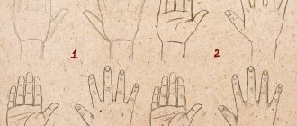
How are muscles different? The photos presented in our article will help us figure this out.
Skeletal muscles are one of the main components of the musculoskeletal system. They allow you to move and maintain balance, and are also involved in the process of breathing, voice production and other functions.
There are more than 600 muscles in the human body. As a percentage, their total mass is 40% of the total body mass. Muscles are classified by shape and structure:
- thick fusiform;
- thin lamellar.
Muscle counting
When most people ask about the exact number of muscles, they are talking about the major skeletal ones - pectorals, deltoids, glutes, biceps and triceps. However, there are hundreds of other small muscles in the human body located in the arms, legs and face.
An approximate figure is about 700 skeletal muscles. This number also includes about 400 muscles, the existence of which is mainly known only to specialists. They are located around the tongue and vocal apparatus, the eyeball or the pelvic floor.
Classification makes learning easier
The division of skeletal muscles into groups is carried out depending on their location and significance in the activity of various organs of the body. Main groups:
Muscles of the head and neck:
- facial expressions - are used when smiling, communicating and creating various grimaces, while ensuring the movement of the constituent parts of the face;
- chewing – promote a change in the position of the maxillofacial region;
- voluntary muscles of the internal organs of the head (soft palate, tongue, eyes, middle ear).
Skeletal muscle groups of the cervical spine:
- superficial – promote inclined and rotational movements of the head;
- middle - create the lower wall of the oral cavity and promote downward movement of the jaw, hyoid bone and laryngeal cartilages;
- deep ones tilt and turn the head, create elevation of the first and second ribs.
The muscles, photos of which you see here, are responsible for the torso and are divided into muscle bundles of the following sections:
- thoracic – activates the upper torso and arms, and also helps to change the position of the ribs during breathing;
- abdominal section - allows blood to move through the veins, changes the position of the chest during breathing, affects the functioning of the intestinal tract, promotes flexion of the torso;
- dorsal - creates the motor system of the upper limbs.
Muscles of the limbs:
- upper - consist of muscle tissue of the shoulder girdle and free upper limb, help move the arm in the shoulder joint capsule and create movements of the wrist and fingers;
- lower - play a major role in the movement of a person in space, are divided into the muscles of the pelvic girdle and the free part.
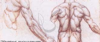
Superficial back muscles
Superficial muscles are represented by:
- The trapezius muscle, attached to all the vertebrae of the thoracic region and its second end to the clavicular bone and scapular spine, is responsible for flexing the head. It is responsible for the movement of the scapula. The upper part raises and the lower part lowers. When you move your arms back, the middle part of the muscle brings your shoulder blades closer to your spine. Also attached to the base of the skull and neck.
- The latissimus dorsi muscle, following the trapezius, is attached to all other parts of the lower spine and to the vertebrae of the anterior chest, thus covering the entire torso in a full rotation. It is not only a corset for the human body, but also pulls the shoulders and arms back, while simultaneously turning them inward. It is one of those that belong to the “large muscles” group, since it is one of the largest in the entire body.
- The rhomboid muscles, both large and small, lie under the trapezius and are attached with their bundles to the lower cervical and capture 4 vertebrae of the thoracic region, and with the other end they are attached to the bone of the scapula and are responsible for its approach to the center.
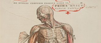
Structure of skeletal muscle
In its structure, it has a huge number of elongated muscle fibers with a diameter of 10 to 100 microns, their length ranges from 1 to 12 cm. The fibers (microfibrils) are thin - actin, and thick - myosin.
The former consist of a protein that has a fibrillar structure. It's called actin. Thick fibers are composed of different types of myosin. They differ in the time it takes to decompose the ATP molecule, which causes different contraction rates.
Myosin in smooth muscle cells is dispersed, although there is a large amount of protein, which, in turn, is significant in prolonged tonic contraction.
The structure of skeletal muscle is similar to a rope or stranded wire woven from fibers. It is surrounded on top by a thin sheath of connective tissue called the epimysium. From its inner surface, deeper into the muscle, thinner branches of connective tissue extend, creating septa. They are “wrapped” with individual bundles of muscle tissue, which contain up to 100 fibrils in each. Narrower branches extend from them even deeper.
The circulatory and nervous systems penetrate through all layers into the skeletal muscles. The arterial vein runs along the perimysium - this is the connective tissue covering the bundles of muscle fibers. Arterial and venous capillaries are located nearby.
Muscle classification
The human muscular system is characterized by a variety of muscle shapes, which in turn are divided into simple and complex. Simple: spindle-shaped, straight, long, short, wide. Complex muscles include the multicipital muscles. As we have already said, if the muscles have a common tendon, and there are two or more heads, then they are called biceps (biceps), triceps (triceps) or quadriceps (quadriceps), and multitendon and digastric muscles are also multi-headed. The following types of muscles with a certain geometric shape are also complex: quadrate, deltoid, soleus, pyramidal, round, serrated, triangular, rhomboid, soleus.
The main functions of muscles are flexion, extension, abduction, adduction, supination, pronation, elevation, descent, straightening and more. The term supination means outward rotation, and the term pronation means inward rotation.
According to the direction of the fibers, muscles are divided into: straight, transverse, circular, oblique, unipennate, bipennate, multipennate, semitendinosus and semimembranosus.
In relation to joints , taking into account the number of joints through which they are thrown: single-joint, double-joint and multi-joint.
Development process
Skeletal muscles develop from the mesoderm. Somites are formed on the side of the neural groove. After time, myotomes are released into them. Their cells, taking on a spindle shape, evolve into myoblasts, which divide. Some of them progress, while others remain unchanged and form myosatellite cells.
A small part of myoblasts, due to the contact of the poles, creates contact with each other, then the plasma membranes disintegrate in the contact zone. Thanks to the fusion of cells, symplasts are created. Undifferentiated young muscle cells move to them, being in the same environment with the myosymplast of the basement membrane.
Facial views
If we talk about how many muscles a person has on his face, the answer can be found in anatomy textbooks. But even there the data is ambiguous, the number is 56-67. They differ from other muscles in the way they are attached. Some of them are adjacent to the bones, while others are adjacent to the mucous membrane or skin.
They can be divided into:
- Muscles with which a person can grind food. These are the muscles that move the lower jaw.
- Facial expressions that move a person’s face. Thanks to them, a person easily expresses his emotional state.
Even though the chewing varieties are some of the strongest, they are quite small in size.
This is interesting! What does the structure and diagram of human skin look like?
Functions of skeletal muscles
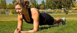
This muscle is the basis of the musculoskeletal system. If it is strong, it is easier to maintain the body in the desired position, and the likelihood of stooping or scoliosis is minimized. Everyone knows about the benefits of playing sports, so let’s look at the role that muscles play in this.
The contractile tissue of skeletal muscles performs many different functions in the human body that are necessary for the correct positioning of the body and the interaction of its individual parts with each other.
Muscles perform the following functions:
- create body mobility;
- protect the thermal energy created inside the body;
- promote movement and vertical retention in space;
- promote contraction of the airways and help with swallowing;
- form facial expressions;
- promote heat production.
Types of muscles
There are three types of muscles in the human body:
- skeletal (they are also called striated);
- smooth;
- and myocardium, or heart muscle.
Smooth muscles form the walls of internal organs and blood vessels. Their distinctive feature is that they work independently of a person’s consciousness: it is impossible to stop, for example, peristalsis (contractions) of the intestines by force of will. The movements of such muscles are slow and monotonous, but they work continuously, without rest, throughout their lives.
Skeletal muscles are responsible for maintaining the body in balance and performing a variety of movements. Do you feel like you are “just” sitting in a chair and relaxing? In fact, dozens of your skeletal muscles are working during this time. The work of skeletal muscles can be controlled by willpower. Striated muscles are capable of contracting quickly and relaxing just as quickly, but intense activity leads to fatigue relatively quickly.
Cardiac muscle uniquely combines the qualities of skeletal and smooth muscles. Just like skeletal muscles, the myocardium is capable of working intensively and contracting rapidly. Just like smooth muscles, it is practically tireless and does not depend on a person’s volitional effort.
By the way, strength training not only “sculpts the relief” and increases the strength of our skeletal muscles - it also indirectly improves the quality of work of smooth muscles and cardiac muscle. By the way, this will also lead to a “feedback” effect - the strengthened heart muscle, developed through endurance training, works more intensely and efficiently, which is expressed in improved blood supply to the entire body, including skeletal muscles, which thanks to this can endure even greater loads. Trained, developed skeletal muscles form a powerful “corset” that supports internal organs, which plays an important role in normalizing digestive processes. Normal digestion, in turn, means normal nutrition of all organs of the body, and muscles in particular.
Different types of muscles differ in their structure, but we will take a closer look at the structure of skeletal muscle, as it is directly related to the process of strength training.
Ongoing support
When muscle tissue is at rest, there is always a slight tension in it, called muscle tone. It is formed due to minor impulse frequencies that enter the muscles from the spinal cord. Their action is determined by signals penetrating from the head to the spinal motor neurons. Muscle tone also depends on their general condition:
- sprains;
- level of filling of muscle cases;
- blood enrichment;
- general water and salt balance.
A person has the ability to regulate the level of muscle load. As a result of prolonged physical exercise or severe emotional and nervous stress, muscle tone involuntarily increases.
Some interesting facts
- Everyday walking at a measured pace, which we are taught to do in early childhood, uses more than 200 muscles, smiling activates 17 muscles, crying - about 34.
- An adult man of athletic build can weigh an order of magnitude more than a full man. This is explained by the anatomy of human muscles, the density of which is much greater than the density of fat.
- Most people consider the tongue to be the strongest muscle in humans, but this is not entirely true. After research, scientists came to the conclusion that the tongue consists of several muscles and it is not entirely correct to consider it as a whole. Therefore, the strongest include the chewing muscles, which can create a load of up to 100 kg, the gluteal and calf muscles.
- The most enduring muscle is rightfully the heart muscle, which does not stop for a second throughout human life. With age, the human heart becomes depleted due to the short-lived supply of glycogen in the muscle walls and an excess of calcium in the structure, which leads to flabbiness and decreased tone.
- In the absence of periodic physical activity, total muscle mass can be burned, like a fat layer. This problem becomes relevant after 40 years, when this process reaches 2-3% per year, after 60 - up to 5%. This is why physical activity is a must in middle age to maintain muscle tone and overall fitness.
- According to foreign scientists, dark chocolate is one of the best sources of fuel for muscle tissue, including the heart and brain. The daily norm for an adult, physically strong person is 40-50 grams per day (a quarter of an ordinary tile). Among other things, chocolate is a source of endorphin (the hormone of joy), which, in addition to physical condition, allows you to maintain optimal morale.
- The palmaris longus muscle, responsible for the release of claws in animals, is found in only 15% of the population and serves as the main source of fibers when needed.
- Exhausting physical activity can raise the core temperature of a person’s body by up to 40 degrees. During training, microtears of muscle tissue occur, the complete recovery of which requires at least 48 hours, with the triceps recovering fastest, and the muscles of the back and legs slower.
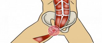
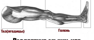
- The longest muscle in the body is the sartorius, which belongs to the anterior group of the thigh; the average length in an adult is 43 cm. It is responsible for the process of throwing one leg over the other. The shortest one is the stirrup, located in the ear, its length is only 1.27 mm. The main function is the movement of the eardrum. Ozoceae family, read our article.
Let's look at this process in more detail.
First, the brain sends an impulse through a system of neurons, which reaches the motor neuron adjacent to the muscle bundle. Next, the efferent neuron is innervated from the synoptic vesicle, and a neurotransmitter is released. It binds to receptors on the sarcolemma of the muscle fiber and opens a sodium channel, which leads to depolarization of the membrane, causing an action potential. When present in sufficient quantities, the neurotransmitter stimulates the production of calcium ions. It then binds to troponin and stimulates its contraction. This, in turn, pulls back tropomeasesin, allowing actin to combine with myosin.
Next, the process of actin filament sliding relative to the myosin filament begins, resulting in skeletal muscle contraction. A schematic diagram will help you understand the process of compression of striated muscle bundles.
A little anatomy
The longissimus dorsi muscles are one of the key muscles in exercises for flexion and extension of the torso. They are located along the entire length of the back, being close to the spinal column. The longissimus muscles are attached to the spine, sacrum and base of the skull with the help of tendons. You can activate their work using exercises such as hyperextension, deadlifts and similar ones described below.
In addition, the longissimus is surrounded by a number of other important muscles, which include the latissimus and teres major. The lats are used to a greater extent during pull-ups and bent-over barbell rows and are located in the lumbar area. The teres major muscles are located towards the middle of the back and are activated through similar exercises.
Next, let's move on to a description of exercises that can strengthen, as well as increase the strength and volume of the longissimus dorsi muscles.
How skeletal muscles work
The interaction of a large number of muscle bundles contributes to various movements of the body.
The work of skeletal muscles can occur in the following ways:
- synergistic muscles work in one direction;
- Antagonist muscles promote opposite movements to produce tension.
The antagonistic action of muscles is one of the main factors in the activity of the musculoskeletal system. When performing any action, not only the muscle fibers that perform it, but also their antagonists are included in the work. They promote counteraction and give the movement concreteness and grace.
When acting on a joint, striated skeletal muscle performs complex work. Its character is determined by the location of the joint axis and the relative position of the muscle.
Some functions of skeletal muscle are poorly understood and often not discussed. For example, some of the bundles act as a lever for the operation of the bones of the skeleton.
Pectoral muscles
The muscles of the thoracic region can be divided into two groups, which include the upper limbs and shoulder girdle muscles:
- The pectoralis major muscle is the uppermost muscle, triangular in shape and starting from the clavicle bone near the shoulder, joining the sternum from the 2nd to 7th rib. The pectoralis major muscle is responsible for moving the arm forward and inward and is also involved in raising the ribs during inhalation.
- The pectoralis minor muscle is located somewhat deeper and is attached at one end to the scapula and the other to the ribs, from the 2nd to the 5th. Participates in its forward and downward movement and, like the large one, is a levator of the ribs during inspiration.
- Another representative of the small muscles is the subclavian muscle. It is stretched between the collarbone and the upper right rib. Pulls it downward, thus fixing and holding it.
- The serratus anterior muscle wraps around the lateral surface of the chest. One end is attached to the 9th rib, and the other to the lower corner of the edge of the scapula. Pulls it forward, rotates it. This is necessary to move the arm above the horizontal position. Also, in collaboration with the rhomboid muscle, it presses the shoulder blade tightly to the body.
Muscle work at the cellular level
The action of skeletal muscles is carried out by two proteins: actin and myosin. These components have the ability to move relative to each other.
For muscle tissue to work, it is necessary to consume energy contained in the chemical bonds of organic compounds. The breakdown and oxidation of such substances occurs in the muscles. There is always air present here, and energy is released, 33% of all this is spent on the performance of muscle tissue, and 67% is transferred to other tissues and spent on maintaining a constant body temperature.
Breathing muscles
The muscles of the trunk also include those involved in breathing. The external and internal intercostal muscles are located in the space between the ribs and are the main participants during inhalation and exhalation.
The diaphragm is the most unusually located flat muscle, which has a dome-shaped appearance. It is directed with the convex part upward. In its action it is a piston pump to carry out the breathing function. It is this muscle that compresses and unclenches the lungs, forcing them to fill with air and releasing it. The diaphragm is attached around the entire perimeter of the chest. It is stretched over the ribs, spine, and lower chest.
Diseases of the skeletal muscles
In most cases, deviations from the norm in muscle functioning are due to the pathological state of the responsible parts of the nervous system.
The most common pathologies of skeletal muscles:
- Muscle cramps are an imbalance of electrolyte in the extracellular fluid surrounding muscle and nerve fibers, as well as changes in osmotic pressure in it, especially its increase.
- Hypocalcemic tetany is an involuntary tetanic contraction of skeletal muscle observed when the extracellular Ca2+ concentration falls to approximately 40% of normal levels.
- Muscular dystrophy is characterized by progressive degeneration of skeletal muscle fibers and myocardium, as well as muscle disability, which can lead to death due to respiratory or cardiac failure.
- Myasthenia gravis is a chronic autoimmune disease in which antibodies to the nicotinic ACh receptor are formed in the body.
How many muscles are in the human body
Human muscles form a complex system. They differ from each other in size, functions, and location. It is generally accepted that there are 640 muscles in the body. These include smooth, skeletal and cardiac. But according to some estimates there may be up to 850.
Muscle names
The name of the muscles reflects either their appearance - latissimus, rectus, or location - sternocleidomastoid.
Many of them are named according to the functions they perform - extensor finger.
Some names have been preserved since the Middle Ages, for example, the sartorius muscle is the one that is involved in flexing the hip, this is the position in which tailors sat at the machine.
Often the name also reflects the location.
According to localization, several groups are distinguished: muscles of the head, neck, torso, upper extremities, lower extremities. Not all of them participate in physical activity.
But you need to know the location of the most famous muscles that are most often involved in training.
Let's take a look at the main muscles in our body that we strive to transform more than others through training and nutrition:
- Trapezoid (Trapezius).
- Deltoid.
- Biceps.
- Rhomboid.
- Large round (Teres major).
- Triceps.
- Extensor carpi radialis (Extensor carpi radialis).
- Extensor digititi minimi.
- Extensor carpi ulnaris (Extensor carpi ulnaris).
- Latissimus dorsi muscle.
- Extensor digitorum.
- Serratus anterior muscle.
- Rectus abdominis muscle.
- External oblique abdominal muscle.
- Lumbar fascia (Thoracolumbar fascia).
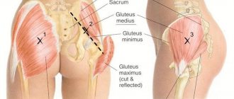
- Gluteus maximus muscle (Gluteus maximus).
- Long adductor muscle (Adductor longus).
- Gracilis muscle of the thigh.
- Vastus lateralis muscle.
- Vastus medialis (vastus medialis).
- Semimembranosus femoris muscle.
- Tibialis anterior muscle.
- Semitendinosus muscle.
- Peroneus longus muscle (Peroneus longus).
- Biceps femoris (biceps femoris).
- Calf muscle (Gastrocnemius).
- Soleus muscle.
- Extensor hallucis brevis (extensor hallucis brevis).
- Short extensor of the toes (Extensor digitorum brevis).
- Sartorius muscle.
- Pectineus muscle.
- Rectus femoris muscle.
- Tensor fasciae latae.
- Gluteus medius (gluteus medius).
- Palmaris longus (Palmaris longus).
- Extensor carpi radialis (Flexor carpi radialis).
- Brachioradialis muscle.
- Pectoralis major muscle.
- Sternocleidomastoid muscle (Sternocleidomastoideus).
Relaxation and restoration of skeletal muscles
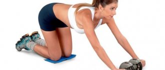
Proper nutrition, lifestyle and regular exercise will help you become the owner of healthy and beautiful skeletal muscles. You don't have to lift weights and build muscle. Regular cardio training and yoga are enough.
Do not forget about the mandatory intake of essential vitamins and minerals, as well as regular visits to saunas and baths with brooms, which allow you to enrich muscle tissue and blood vessels with oxygen.
Systematic relaxing massages will increase the elasticity and reproduction of muscle bundles. Visiting a cryosauna also has a positive effect on the structure and functioning of skeletal muscles.
Functions of human muscles
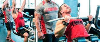
Every athlete who wants to build muscles and change the shape of the body must know their anatomy and functions. You need to understand what exercises you need to do, how to increase the working weights in the exercises. There are several muscles that are most often involved in training.
Neck
You can pump up the sternocleidomastoid muscle from the neck muscles. She is responsible for tilting the head in all directions, as well as turning. Strengthening it is important for those athletes who play football, boxing, and wrestling.
You can do exercises with weights.
Torso
From the torso, special attention is paid to the abdomen, back, pectoral muscles, and neck.
- The pectoralis major is responsible for adducting the upper limbs, lifting up, and lowering. You need to do push-ups from the floor or parallel bars, abducting your arms on a block, and chest press. By the way, I have an article about how to pump up your breasts at home.
- The rectus abdominis muscle is responsible for bending the torso forward. A beautiful relief can be created by performing crunches from a lying position. I advise you to read my article about how to quickly pump up your abs at home.
- The external obliques help with forward bending and also perform lateral bending. They train while throwing the javelin, playing tennis, performing side bends and twisting.
- Trapezius - it helps lift the shoulders, move the shoulder blades, as well as the head back and forth and to the sides. Trained by weightlifters, gymnasts, during rowing and while pressing. Here is an article about how to pump up your trapezius.
- Latissimus – bending the torso to the sides, moving the arms back. Works during rowing, gymnastics and weightlifting. You can train using pull-ups on the bar. Read the link in detail about how to pump up your back.
Upper limbs
Mostly men try to pump up their arm muscles, but women will also find it useful to know the following information. To create a beautiful relief, you will need to work on the following types of muscles of the upper limbs:
- Biceps (biceps) – bending at the elbows, turning the wrist. Train for any exercises that include arm curls, as well as during rowing. Here is an article about how to pump up your arms.
- The coracobrachialis is responsible for raising the arms. You can train during bowling, arm wrestling, javelin throwing.
- Brachial – adduction of the forearm. To train it, you need to row, climb a rope, and perform weighted arm curls. Here is a detailed article about how to pump up your forearms.
- The triceps (triceps) is responsible for moving the upper limbs back. You need to do handstands and exercises involving extension of your arms.
- The deltoids are responsible for lifting the upper limbs. They train during gymnastics, weightlifting, and throwing. You can also do presses and lifting weights. Read the article about how to pump up deltoids.
Lower limbs
It is easier to train the leg muscles; there are many sports that put stress on the lower limbs.
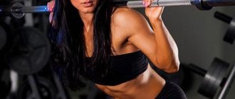
- The quadriceps is responsible for rotation and supination, straightening in the hip joint. All types of squats, presses, leg extensions with weights are useful. He also trains for cycling, football, and athletics. Here is an article about how to pump your legs.
- Biceps hamstrings – for leg curls. To pump up, you need to do any exercises related to this movement. The most effective exercise for the hamstrings is the barbell deadlift.
- The gluteus maximus rotates the hip. Swimming, skiing, cycling are useful. Read the article about how to quickly pump up your buttocks.
- The gastrocnemius is involved in the work of the knee joint and the rotation of the foot. Half squats, jumping, running, cycling are useful.
- The soleus extends the foot. Trains with calf raises.
- The tibia and fibula are involved in turning and other movements of the foot. You need to do a calf raise.
Bent-overs with a barbell
When bending over with a barbell on your shoulders, the longissimus muscles act as the most important muscles. During execution, the knees should be slightly bent, and the bends should be done until the torso is almost parallel to the floor.
The key nuance of this exercise is that the weight of the bar along with the weights should not be too large, since this will reduce the emphasis on the longissimus muscles and the entire load will be transferred to the posterior muscles of the thighs.
The principle of performing tilts with a barbell on your shoulders is as follows: you need to perform 4 sets of 10 repetitions each.
If you achieve good results in bending over with a barbell, you can switch to the same bending, but only in a sitting position. The weight of the projectile in this case should be slightly less, and the volume of training will increase to 15 repetitions in 5 approaches.