Good afternoon, dear readers. The good news is that the simulation of distance learning at my university is nearing its end, which means that you and I have time to study fundamental medicine. Today we will look at the deep muscles of the neck.
This is a topic that is often ignored by both students and even teachers. All anatomy buffs know the superficial and middle muscles of the neck, just like the well-known triangles. But there are always problems with deep muscles.
This is partly due to the muscles in the back - parts of some of them are located in the neck, and this leads to serious confusion. Let's try to figure this out and start, as always, with classification.
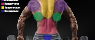
The deep muscles of the neck are divided into three groups. These are the lateral muscles, the median muscles and the suboccipital muscles. Classification here, as in all anatomy in general, is a great way to remember the topic, because these three groups reflect the location of the muscles.
Lateral group
This is a very simple group of neck muscles, and it would be most logical to start with it. The lateral muscles of the neck are located directly on the side, that is, laterally. By the way, if you are confused about anatomical terminology, be sure to look here.
So, imagine that the corpse was sawed in the frontal plane (that is, along the blue rectangle).
In this cut we look at the preparation from the face:
This will be our main cut in this tutorial. By the way, this is from Sinelnikov’s atlas. Let's highlight the lateral muscle group in this cut, which we will now work with. I highlighted the lateral group of muscles only on one side, because more muscles were prepared on the other side and we can not see all the muscles of the lateral group.
Please note that in this preparation almost the entire skull has been sawed off, except for the back of the occipital and temporal bones, and all the muscles of the neck of the superficial and middle layers have been completely removed.
The lateral group of deep neck muscles is represented by three scalene muscles. These are paired muscles (although all the muscles of the neck are paired, except for the subcutaneous muscle).
Anterior scalene muscle (musculus scalenus anterior)
This muscle is indeed located closer to the anterior surface of the body than the other scalene muscles. By this sign alone we can easily detect the anterior scalene muscle:
If we look at the familiar tablet of the superficial muscles of the neck, we can even see the anterior scalene muscle there. You can see how it is covered by the sternocleidomastoid muscle (11) in front and the levator scapula muscle (19) in the back.
In the last picture, as in the cadaveric material, you can only see the lower part of the belly of the anterior scalene muscle. From this angle it is quite difficult to understand the shape of this muscle. However, the anterior scalene muscle has a very interesting shape. The anterior scalene muscle is similar to a tree branch, with three smaller branches extending from a large trunk. This can be clearly seen in the picture, which shows only the anterior scalene muscle:
Beginning: anterior tubercles of 3-6 cervical vertebrae;
Insertion: tubercle of scalene muscle of 1st rib;
Function: with a fixed spine, pulls the first rib upward; with a fixed chest, tilts the cervical spine forward.
Middle scalene muscle (musculus scalenus medius)
Let's move a little towards the back of the head from the anterior scalene muscle. Here we will see the middle scalene muscle - larger and more powerful than the anterior muscle. Let's mark it in blue on the same tablet on which we marked the anterior scalene muscle. The middle scalene muscle looks small here due to the fact that it is covered by the anterior and posterior scalene muscles:
We select the middle scalene muscle and on the tablet with the superficial and middle muscles:
And now let's look at the scalene medius muscle, in an illustration where all the other muscles are missing:
As you can see, it looks more like a hand with long, branchy fingers than a tree branch. In the X-Men comics, there is a lady supervillain named Lady Deathstrike, she is one of Wolverine's arch-enemies. I remember this formidable aunt when I look at the middle scalene muscle.
Origin: anterior tubercles of the first six cervical vertebrae. There are more insertion points and therefore more "fingers" than the anterior muscle;
Attachment: first rib - that is, the same as the previous muscle, only slightly behind it;
Function: similar to the functions of the anterior scalene muscle.
Stair triangle
American atlases and anatomy textbooks always indicate the space bounded anteriorly by the rectus scalene muscle, posteriorly by the middle scalene muscle, and inferiorly by the first rib. This space has the form of a triangle and is therefore called a ladder triangle (scalene triangle).
This is a very important topographic formation, because the subclavian artery and nerve trunks that form the brachial plexus pass through this triangle. All of the above can be clearly seen in this illustration.
Let's tint all these anatomical formations for greater convenience. As in the illustrations above, I highlighted the anterior scalene muscle in yellow, the posterior scalene muscle in blue, and the rib in green.
Still looks too confusing? Let's select our triangle:
By the way, we can also highlight the scalene triangle on the preparation we are working with in this lesson:
Posterior scalene muscle (musculus scalenus posterior)
We have come to the last muscle of the lateral group, the posterior scalene muscle. Let's see how it looks on our main tablet (I've highlighted it in green):
On our tablet with the superficial and middle muscles of the neck, we can see a very small section of the posterior scalene muscle:
In the illustration, where we see only this muscle without all the others, we can notice its special features. This is a muscle with a fairly wide belly, which starts from three vertebrae and forms a “branch” of a tree, like the anterior scalene muscle. However, its fundamental difference from the first two muscles is that it is attached to the second rib, and not to the first.
I also found an illustration from my favorite atlas by Yu.L. The golden area is where we can see the posterior scalene muscle. Here, all the back muscles and suboccipital muscles of the neck were removed (we will learn them later), as well as the vertebral arches were sawed off and the spinal canal was opened. There is a lot of interesting stuff in this illustration, but it is the posterior scalene muscle that interests us. I'm quite sensitive to the idea of highlighting anything in the illustrations from Zolotko's atlas (it's too beautiful), so I just emphasized the name of the posterior scalene muscle:
Beginning: posterior tubercles of the 5th, 6th and 7th cervical vertebrae;
Attachment: outer surface of 2 ribs;
Function: raising the 2nd rib with a strengthened spinal column. With a strengthened chest and bilateral contraction, the posterior scalene muscle tilts the cervical spine forward.
The structure of muscle fiber and the mechanism of muscle function
Each muscle has approximately the same internal structure: on the outside, the multinucleated cell is covered with sarcolemma, which is a special membrane substance filled with connective tissue. On the inside, it is a set of muscle bundles of different orders that are united through the endomysium - connective tissue.
At the same time, each muscle contains a series of blood vessels and capillaries to ensure sufficient oxygen supply during exercise. The nerves that penetrate the fibers provide conductivity, excitability and quick, high-quality response during work.
Muscle cells have several nuclei, since during active work they are able to generate thermal energy thanks to numerous mitochondria.
The most important functions of muscle fibers are contractility and excitability, which are ensured by the joint interaction of nerve and protein structures and are controlled by the central nervous system (brain and spinal cord). Muscles owe their contractile abilities to special proteins: actin and myosin. Muscle fibers have a diameter of 10 to 80 micrometers and a length of up to 35 cm.
The main types of skeletal muscle fibers are:
- Slow (ST or type I). Slow twitch fibers can sustain contractions for long periods of time and are also highly resistant to fatigue.
- Fast (FT or type II). Fast twitch fibers create short, powerful contractions.
Middle group
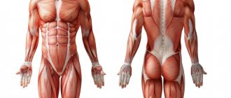
The medial group of neck muscles includes two muscles: the longus colli muscle and the longus capitis muscle. The fundamental point here is the difference from the longissimus dorsi muscle - they are often confused, because both the shape and the name are similar. It is necessary to understand that the longissimus muscle is located on the dorsal side of the vertebrae, that is, on the back side. And the long muscles are located on the ventral side. In this picture I have shown the approximate location of the longissimus muscles (neck muscles) in red and the longissimus muscles (back muscles) in green.
Now let's look directly at the muscles of the middle group.
Long colli muscle (musculus longus coli)
As I already said, the most important point is the location of the longus colli muscle just anterior to the spinal column. Knowing just this one point will help you not get confused when working with cadaveric material or complex anatomical plates.
The longus colli muscle is really long, extending from the atlas to the 4th thoracic vertebra. The longus colli muscle is clearly visible in our main preparation:
We can also use an illustration from Frank Netter's atlas, the longus colli muscle is clearly visible here:
Origin: bodies of the first 3 thoracic vertebrae and 2-7 cervical vertebrae;
Attachment: anterior tubercle of the atlas, anterior tubercles of the costotransverse processes of 2-7 cervical vertebrae;
Function: tilts the cervical spine, and, accordingly, the head, down.
Long muscle of the head (musculus longus capitis)
By function, this muscle is a synergist of the longus colli muscle, that is, it performs the same functions, acting together with the longus colli muscle. You can easily avoid the trap with names and confusion with the longissimus capitis muscle if you look at the picture with four skeletons just above.
Here the longissimus capitis muscle is highlighted in yellow (it has been removed to our right):
A very similar illustration by Frank Netter:
Beginning: anterior tubercles of 3, 4, 5, 6 vertebrae;
Insertion: inferior surface of occipital bone;
Function: tilts the cervical spine, and, accordingly, the head, down.
You can also find the middle group neck muscles in a horizontal section. Here we see only the long muscles of the neck, because this is a cut at the level of the 7th cervical vertebra, the rest is located higher. I've highlighted the long neck muscles in green:
The fundamental point is the location of the deep muscles of the neck (the vertebra is highlighted in black):
Human body muscle classification table
The types of muscles in humans are divided into 2 groups:
- Smooth. They are found inside the walls of blood vessels, the uterus, intestinal walls and the internal muscles of the eye, and are controlled by the autonomic nervous system. Smooth muscle helps with everything from blood circulation to digestion.
- Cross-striped. This means that each muscle fiber has stripes or linear markings that can be seen under a microscope. The stripes correspond to sarcomeres that contract rapidly and in a coordinated manner. This group includes:
- Heartfelt. It is present only in the heart and is associated with the autonomic nervous system. Unlike skeletal fibers, cardiac muscle fibers are arranged in a branching rather than a linear pattern. Its main properties are endurance and consistency.
- Skeletal muscles. They are controlled by the somatic nervous system. Most muscles belong to this group. They are attached to the bones, and sometimes, like facial ones, to the skin.
Types of muscles in humans. Titles
Muscles have different shapes, development, structure, and perform various functions. According to their location, muscles are deep, located closer to the skeleton, and superficial, located closer to the skin.
| Structure | Simple: spindle-shaped, long, short, wide. |
| Complex: 2-headed, 3-headed, 4-headed, multi-headed, vubdominal, multitendinous. | |
| Complex: square, trapezoidal, diamond-shaped, deltoid, jagged, round, soleus, triangular, pyramidal. | |
| Nature of movements | Flexion, extension, abduction, drive, rotation, lifting, lowering, straightening, dilators, sphincters, antagonists that perform opposite movements, synergists that perform movements in a friendly manner. |
| Joint involvement | 1-joint, 2-joint, 3-joint. |
| Fiber direction | Straight, circular, transverse, oblique (unipinnate, 2-pinnate, multipinnate, semimembranosus, semitendinosus). |
The thickened part of the muscle tissue is called the abdomen, and the final sections are called the head, which is attached to the skeleton. There are muscles with several bellies and heads.
Suboccipital muscles
This is the last and deepest layer of neck muscles. In truth, in most textbooks the suboccipital muscles are not included in the group of deep muscles; they are an independent, fourth group of neck muscles. That is, they are too deep to be classified as simply deep muscles. Let's look at these muscles.
Anterior rectus capitis muscle (musculus rectus capitis anterior)
The rectus capitis anterior muscle is a very small and short muscle that looks like a small rectangle connecting the first cervical vertebra and the skull. It is located the deepest, so it will be convenient for us to view it from the front, from the already familiar cut of the neck in the frontal plane:
I am always confused by Latin terms that contradict the appearance of the object being called. For example, this rectus muscle is not completely straight. However, if you remember that it is located precisely in the inner part of the neck, you will immediately detect it.
Here the rectus capitis muscle is also tinted from the same angle (this is Gray's atlas). Unfortunately, not all the muscle has been removed from the half to our left, and we can see the rectus capitis muscle only to our right.
Origin: lateral masses of the atlas;
Insertion: inferior surface of the occipital bone, anterior to the anterior margin of the foramen magnum;
Function: with unilateral contraction, it tilts the head in its direction; with bilateral contraction, both muscles tilt the head forward.
Lateral rectus capitis muscle (musculus rectus capitis lateralis)
If it all ended on this muscle, it would be very cool, wouldn't it? We move slightly to the lateral side from the anterior rectus capitis muscle and get the lateral rectus capitis muscle. These two muscles form an acute angle - very convenient for memorization.
Let's look at the same muscle in Gray's atlas:
Origin: anterior part of the transverse process of the atlas;
Insertion: jugular process of occipital bone;
Function: with unilateral contraction, it tilts the head in its direction; with bilateral contraction, both muscles tilt the head forward.
Major posterior rectus capitis muscle (musculus rectus capitis posterior major)
From now on, we can say goodbye to the usual cutting in the frontal plane. Now we need to look at the back of the neck area. This angle will suit us:
As you can see, this is the atlanto-occipital joint, the upper part of the cervical spine and the back of the skull. We will work with this tablet.
So, here we see several muscles, and we are interested in the largest one. This is the rectus capitis posterior major muscle. There are quite a lot of muscles here, but you can remember that the BZPMG is the largest of all the suboccipital muscles. I've highlighted the rectus capitis posterior major muscle in yellow:
Also, on the Internet, I found another cool illustration - it looks like Grant’s atlas, but I could be wrong. Here we can also see the large posterior rectus capitis muscle, it is designated by the letters MA:
And another illustration from Gray’s atlas:
Origin: spinous process of the axis (2nd cervical vertebra);
Insertion: inferior nuchal line, lateral margin;
Function: throwing the head back, straightening the head from a position where the chin is pressed to the chest.
Rectus capitis posterior minor (musculus rectus capitis posterior minor)
We move a little medial to the previous muscle and see a muscle that is smaller in size, but very similar in shape. This is the rectus capitis posterior minor muscle. Probably, of all the “rectus” muscles, this muscle is the most straight.
On our tablet we can see both of the posterior rectus minor muscles. The previous muscle, the BZPMG, is highlighted in yellow (on the other side it has been removed), but I highlighted both MZPMGs in blue:
On our second tablet we can see this muscle, indicated by the letters mi. Although it can be recognized without any markings, it is located the most medial and most vertical of all the muscles of this group:
And a very small illustration from Gray’s atlas:
Origin: posterior tubercle of the atlas;
Insertion: medial part of the inferior nuchal line of the occipital bone;
Function: throwing the head back, straightening the head from a position where the chin is pressed to the chest.
Superior oblique muscle of the head (musculus obliquus capitis superior)
The suboccipital muscles are quite easy to navigate, actually. The superior oblique is very visible on any suboccipital plate. Let's mark it on our tablet in green:
On our other tablet, I also highlighted the superior oblique muscle in green:
In Gray's atlas we can also see the superior oblique capitis muscle:
Origin: transverse process of the atlas;
Attachment: lateral segments of the superior nuchal line;
Function: throwing the head back, straightening the head from a position where the chin is pressed to the chest.
Inferior oblique muscle of the head (musculus obliquus capitis inferior)
This is the last muscle of the entire group. You can easily identify the inferior oblique muscle because this muscle forms an obtuse (almost right) angle with the superior oblique muscle. This angle is always clearly visible. Look, I've added a light orange outline of the inferior oblique muscle to our tablet:
We find the same angle on another tablet:
And finally, the inferior oblique muscle of the head in Gray's atlas:
Origin: spinous process of the axis;
Insertion: transverse process of the atlas;
Function: throwing the head back, straightening the head from a position where the chin is pressed to the chest.
You can practice identifying the deep muscles of the neck of the suboccipital group with an illustration from my favorite Zolotko atlas: