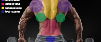
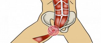
Development of the teres major muscle of the back
Most exercises with dumbbells and pulleys target the lats. This area responds well to load, so progress is noticeable quite quickly. Pumped up lats provide beautiful relief and power to the back. The teres major muscle is located under the latissimus, and it is this muscle that forms the correct position of the spine. Exercises for this area will help:
- make your back wider;
- draw the relief of the “small wing”;
- increase shoulder volume;
- strengthen your hands.
In fact, the teres major muscle is the framework of the latissimus, so without its development it will not be possible to form a truly powerful and sculpted back. This area is responsible for pulling the arm down and back and bringing it toward the body, so developing it will definitely have a positive effect on arm strength. Another important role is to support the spine, so it is recommended to perform such exercises for the back with osteochondrosis.
Infraspinatus stretch
- The first stretching exercise: you need to place your hand behind your back at the level of the lower back, then with the other hand gently pull from the back and up. You must hold your hand in this position for twenty seconds.
- The second stretching exercise: you need to try to reach the opposite shoulder blade with your fingers.
- Third stretching exercise: you need to extend your arm in front of your chest and use your other hand to guide it towards the other half of your chest. Just do this without much tension. The hand is held in this position for twenty seconds.
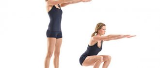
Bodyweight exercises
It is recommended to start by doing simple exercises with your own weight. They have a number of undoubted advantages:
- easy to learn;
- no auxiliary equipment needed;
- can be performed anywhere;
- effectively develop overall endurance and strength.
Let's take a closer look at how to pump up the teres major muscle of the back by working out with your own weight.
On the horizontal bar
Few people understand how to pump up the lateral back muscles, but the usual pull-ups are aimed specifically at developing this area. Overall, this is the best basic movement for comprehensive back development. Pull-ups are performed either with a wide grip on straight arms (with the arm fully extended) or with bent arms. The second option additionally engages the rhomboid back muscle.
The technique is very simple: grab the bar so that your arms are bent at a right angle, from this position pull yourself to your chest, and lower smoothly. The secret to the effectiveness of the movement is simple: when lowering your body, you must not lose the correct bend of your arm, that is, do not straighten it completely. Pull-ups are performed with a wide grip, with your hands positioned wider than your shoulders.
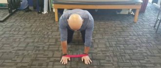
Push ups
Everyone knows that there is nothing better for the chest muscles than push-ups. However, for most people who go to the gym, a surprise is the fact that the classic push-up also uses the deep back muscles. The following techniques for performing the movement will help you make them work.
- Push-ups with wide arms: take a lying position, straighten your arms and place them wider than your shoulders, feet together. Bend your elbows and lower your body until it almost touches the floor, then straighten your arms.
- Diamond push-ups: The hands are positioned so that the thumbs and index fingers of both hands form a triangle.
- With clap: perform a push-up with your arms wide, clapping as you straighten your arms. The exercise is performed with intense pushing of the body from the floor.
Clapping push-ups are a heavier version of the exercise, but this technique also uses the lats.
Plank
The best exercise for strengthening the spine is the plank in various variations. When performing, the rhomboid muscle, which is responsible for posture, is also loaded.
It’s very simple to do: take a lying position, bend your arms and lower yourself onto your forearms. Round your lower back a little, tighten your abs, tighten your buttocks. The exercise is static; you must stand in this position for at least 30 seconds. It is best to perform several approaches of half a minute with a short rest between repetitions.
- Another option is to stand with your arms outstretched. This technique is a little easier, so the time spent in the plank should be increased to one minute or more.
- The plank can be done in motion: lean on your forearms, then straighten your arms, and lower yourself onto your forearms again. Repeat 10-15 times.
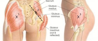
Athletes are often interested in how to pump up the infraspinatus region and oblique muscles of the back. This area is located under the shoulder blade and is worked during the side plank. To take the correct position, you should lean on the forearm of one hand and foot, turning your body and extending your other arm above your head. Stay in this position for half a minute, then repeat on the other side of the body. The movement stretches the oblique muscles of the back and improves posture.
Causes and types of muscle diseases
The main pathology associated with muscle damage includes myositis. Myalgia, that is, muscle pain, is the main symptom of this disease.
This symptom manifests itself as painful nagging or constant aching muscle pain, which is localized both in the projection area of the affected muscle and spreads to neighboring areas (radiates).
Myositis is a disease characterized by inflammatory phenomena of various origins in striated muscles. Not only individual structures are involved in the process, but also entire muscle groups that perform similar functions.
The most common causes of myositis include:
- Injuries of various nature (mainly domestic and industrial).
- Increased physical activity associated with both static stress (long stay in one position) and dynamic loads.
- Infectious diseases caused by bacteria or viruses (acute respiratory viral infections, pneumonia).
- Autoimmune conditions (the human body produces antibodies to the cells of its own tissues, often as a result of past infectious diseases).
- Exposure to toxins (long-term use of alcohol, drugs).
- Exposure to certain groups of drugs that lower blood cholesterol (atorvastatin), immunomodulators (interferons).
Myositis is characterized by the presence of pain that intensifies when moving or feeling the hand. The nature of the pain in this pathology is usually dull or aching and is constant. Also, the upper limb, where the affected muscle is located, becomes less functional.
The teres major muscle is palpated as a painful lump due to tissue swelling caused by a local inflammatory process. If the cause of myositis was an infection suffered by a person, in addition to local inflammatory processes there may also be systemic manifestations of pathology:
- hyperthermia;
- weakness;
- lethargy;
- headache.
According to the duration of its course, myositis can be acute, as well as chronic (can occur in waves).
In order to make a correct diagnosis, it is necessary, first of all, to clarify the history of the disease (localization of pain, its nature, duration, possible causes), the patient’s complaints, conduct a general examination and correctly assess the symptoms based on the information received.
Laboratory (general and biochemical blood tests) and instrumental research methods (ultrasound diagnostics) are auxiliary and allow a more accurate clinical diagnosis based on objective data.
Posture Pulls
A variety of rows is best for strengthening your back muscles. They can be performed with dumbbells, barbells, or on special machines. At home, fitness bands or elastic bands are used.
Rows are easy to learn on your own, especially if you perform the movement on a machine. Light weight exercises can be considered as a way to recover the back from a herniated disc, but only after consulting a doctor.
Horizontal thrust
Performing this movement will strengthen your entire back, relieve muscle tension and effectively work your deltoids and chest. To do this, you need to set the desired weight on the block and attach a V-shaped handle to the carabiner. Grasp the handle, rest your feet on the support and pull the block towards your chest, while simultaneously slightly tilting your body. Then release your efforts and reach for the block, rounding the thoracic spine. Repeat the required number of times.
The movement can be performed with a reverse grip with a standard two-handed handle. In this case, the emphasis shifts to the deep back muscles.
T-bar row
One of the most effective and safe movements, which will allow you to both pump up large muscles and strengthen your deltoids. Working with the T-bar eliminates the help of the body, while the technique is simple. It is necessary to equip the bar with the required weight, rest your feet on special supports, and grab the handle with a direct grip. Lean forward, knees bent. Then raise the bar to waist level and straighten your arms, lowering the weight.
Dumbbell row
The movement is much like the T-bar row. It is necessary to spread your legs, bend your knees, tilt your body forward so that it is parallel to the floor. Take a dumbbell in your hand and use your other hand to lean against the wall. Pull the dumbbell to your waist, then straighten your arm. You need to hold the projectile with your fingers towards you.
Features[edit | edit code]
Infraspinatus muscle
(
m. infraspinatus
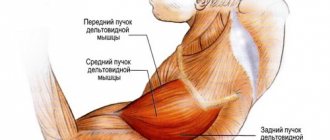
) participates in the formation of the rotator cuff and, weaving into the capsule of the shoulder joint, strengthens it. The infraspinatus muscle is considered a strong external rotator of the shoulder, especially in the final phase, when with external rotation of the greater tuberosity and continued abduction of the arm it is necessary to avoid its impact with the roof of the shoulder joint. The cranial part of this muscle abducts the arm, and the caudal part adducts.
| Functions | Synergists | Antagonists |
| Shoulder external rotation | *M. teres minor *M. deltoideus (spinous part) | *M. pectoralis major *M. deltoideus (clavicular part) *M. latissimus dorsi *M. teres major *M. biceps brachii |
| Shoulder adduction | *M. pectoralis major *M. latissimus dorsi *M. teres major *M. teres minor *M. coracobrachialis *M. biceps brachii (short head) *M. deltoideus (spinous and clavicular parts with the arm already adducted) *M. triceps brachii (long head with arm already abducted) | *M. deltoideus (acromial part) *M. deltoideus (spinous and clavicular parts with the arm abducted) *M. infraspinatus (cranial part) *M. biceps brachii (long head) |
| Shoulder abduction | *M. deltoideus (acromial part) *M. deltoideus (spinous and clavicular parts with the arm abducted) *M. biceps brachii (long head) | *M. pectoralis major *M. latissimus dorsi *M. teres major *M. teres minor *M. coracobrachialis *M. biceps brachii (short head) *M. deltoideus (clavicular and spinous parts with the arm already adducted) *M. infraspinatus (caudal part) *M. triceps brachii (long head with arm already abducted) |
| Shoulder retroversion with the arm abducted | *M. deltoideus (spinous part) *M. teres minor | *M. pectoralis major *M. deltoideus (clavicular part) *M. coracobrachialis *M. biceps brachii (short head) |
Shoulder external rotation. Functional muscle tests[edit | edit code]
Issues and comments
- With this test, it is almost impossible to distinguish between the functions of the teres minor, infraspinatus muscles and the acromial part of the deltoid muscle.
- If the arm is extended, supination at the elbow may appear as external rotation.
- Due to the more superficial location of the latissimus dorsi muscle, it is not always possible to palpate the infraspinatus muscle.
Other exercises
A healthy back is the result of hard work on all muscle groups. To ensure uniform development of this area, you can regularly change the set of exercises, complementing the training plan or changing it. The following movements will help you effectively work your muscles.
- Hyperextension. It is performed in a special simulator for spinal extensors. Lie down on the machine so that your hips rest against the upper roller and place your legs under the lower roller. Crossing your arms behind your head, lower your body down, bending your lower back, then return to the starting position, straightening your spine.
- Bent-overs with a barbell. Stand straight, legs slightly apart, knee soft. Place the barbell on your shoulders, tilt your body forward, arms bent at the elbows, palms holding the bar shoulder-width apart.
- Upper block pull. Sit on a bench, grab the straight handle of the upper block so that your hands are located slightly wider than your shoulders. Pull the chest block while leaning back and bringing your shoulder blades together. Then release the block, moving your body forward, stretching the loaded zones.
Few people know how to pump up the serratus muscles. In fact, they are involved in almost all exercises for developing deltoids and trapezius. For example, simple shrugs with an empty bar will both pump up the serratus muscles and strengthen the entire shoulder girdle. You can supplement your workout with levers, deadlifts, or deadlifts.
Massage of the infraspinatus muscle
As with any other massage, the main goal here is to improve blood circulation and relax the spasming muscle. The simplest, but at the same time quite effective way to massage the infraspinatus muscle is the following. It is performed in a supine position. You need to place a small ball the size of a tennis ball under those points on your back that are most painful, and then relax for a few minutes. When this method is performed correctly, the muscles relax and pain disappears.
Gymnastics for posture
Having figured out how to pump up the round muscles of the back in the gym, you should take a closer look at what you can do for your posture. Exercise therapy (physical therapy) exercises are suitable for performing in case of intervertebral hernia and osteochondrosis. However, you cannot do gymnastics for posture during an exacerbation of the disease. You can perform exercise therapy at home, but only if there is no pain and after the gymnastics complex is approved by your doctor.
If you pump up the diamond-shaped area of the back with osteochondrosis of the cervical spine, you can get rid of tension, dizziness and heaviness after waking up. Osteopaths advise pumping up the longitudinal muscles of the back with instability in the thoracic region and stooping to reduce symptoms. You should figure out how to pump up the oblique muscles for problems in the lumbar region, because they reduce the load on the lumbar region.
For those who don’t know how to pump up their back muscles at home, the following gymnastics complex will come to the rescue.
- Downward-facing dog and up-facing dog are two popular yoga movements. With their help, you can perfectly work out the oblique muscles of the back and the entire spinal region. Movement strengthens the back muscles during intervertebral hernia, protrusion and osteochondrosis. To perform, lie on your stomach on the floor, place your hands in front of you. Straighten your arms, bending at the waist and raising your head - this is the pose of an upward-facing dog. From this position, rise to your feet, without lifting your palms from the floor, lower your head. The body forms a V - this is the downward-facing dog pose. So, changing body position, perform 10 repetitions.
- The bridge is a movement familiar to everyone since childhood, which perfectly stretches the lumbar and thoracic region. Lie on the floor, bend your knees, feet pressed to the floor. Lift your pelvis off the floor, resting on your shoulder blades, elbows and feet. Bend your spine as much as possible and stay in this position for half a minute. Repeat 5 times.
- Boat. This simple movement is often used as a warm-up, as it perfectly prepares the body for the load. You need to lie on your stomach with your arms extended forward. Lift your arms and legs off the floor at the same time, hold the tension for a few seconds, then relax your body. Repeat 10 times.
With these simple movements you can strengthen your back muscles both at home and in the gym. They can be performed as a cool-down after training and as an independent complex for posture. If you have spinal diseases, it is recommended to attend specialized exercise therapy classes at the clinic, or work out in the gym with a rehabilitation specialist. In all other cases, you can master the exercises with the help of training videos.
Treatment of the supraspinatus muscle of the shoulder
The supraspinatus muscle is part of the rotator cuff. This is a group of muscles that provide rotational movements in the joint and maintain its stability. The supraspinatus muscle stabilizes the confluence of the scapula, thereby forming the point of rotation of the shoulder. If it is damaged, lifting the shoulder joint and maintaining it in abduction becomes impossible.
Carpal tunnel syndrome
The distal portion of the supraspinatus muscle plays an important role in the function of the shoulder joint. It is located in the osteofibrous tunnel of the scapula. Normally, the area of the tunnel should correspond to the area of the supraspinatus muscle. If there is a discrepancy, the muscle becomes compressed, which causes pain.
With long-term rotator cuff tunnel syndrome, degeneration of the supraspinatus muscle occurs. Subsequently, its tendon is damaged. To prevent pathological changes, decompression of muscle tissue is required.
Symptoms of this condition:
- chronic pain;
- violation of active movements in the shoulder joint.
X-rays and MRI show an increased size of the acromial end of the clavicle and a degenerative change in the supraspinatus muscle of the shoulder. Treatment for this disease is only surgical. It should be performed as early as possible to avoid significant damage to the tendon.
Stages
At stage 1 (compression stage) of rotator cuff injury, the patient experiences pain when trying to perform full movements in the shoulder joint. The glenohumeral rhythm is maintained. At rest there is no pain or it is mild. There is no pronounced hypotrophy in the supraspinatus fossa of the scapula.
At this stage there are still no significant destructive changes in the supraspinatus muscle. However, X-rays already show pathological changes in the distal end of the clavicle and the acromial clavicular joint. On MRI you can see compression of the supraspinatus muscle in the canal, its funnel-shaped deformation.
At stage 2 (degeneration stage), the strength of the supraspinatus muscle decreases. As a result, there is no adequate stabilization of the humeral head. Dysfunction of the initial stage of arm abduction is noted. The head of the humerus moves upward. Main complaints from the patient:
- inability to fully perform active abduction movements in the shoulder;
- pain at rest, worsening with exercise.
Objectively, there is a violation of the glenohumeral ri). MRI reveals signs of hypotrophy of the supraspinatus muscle. It is compressed by the distal portion of the clavicle. With prolonged untreated syndrome, degenerative processes in the surrounding soft tissues progress.
At stage 3 (anatomical damage stage), pseudoparalysis of the shoulder joint syndrome occurs. Active movements are possible, but they are insignificant in amplitude and are provided mainly by the scapula. External rotation of the shoulder becomes impossible with minimal resistance. Deforming arthrosis of the acromial clavicular joint often develops.
Fresh damage
Treatment results are best for a rupture of the supraspinatus muscle of the shoulder that has occurred recently. In this case, the operation of choice is subacromial decompression of the joint.
The disease is accompanied by chronic pain syndrome, which is associated with inflammation of the supraspinatus muscle of the shoulder. Treatment can be carried out arthroscopically.
This is a minimally invasive operation with a short recovery period and low surgical risks.
Arthroscopic subacromial decompression is performed with the patient lying on the healthy side. The doctor examines the cartilage of the articular surfaces, assesses the condition of the biceps tendon, synovium, and rotator cuff muscles. If degenerative changes are detected, the doctor treats the superficial areas with cutters.
Then the doctor performs an inspection of the subacromial space and excises the subacromial bursa (joint capsule). The entire coracoacromial ligament is removed. Up to 1 cm of the anterioinferior section of the acromion is removed.
To identify and treat possible tears in muscle tissue and areas of fibrosis, the outer surface of the rotator cuff is examined.
The distance from the attachment of the rotator cuff to the greater tubercle of the humerus to the distant edge of the acromion is measured. It should be 3 mm or more.
In this case, there will be enough space for the tendon, and movements in the shoulder will become painless.
After surgery, the drains are removed after 12 hours. Shoulder immobilization lasts up to 2 weeks. It is advisable to begin restoration measures already on the second day. First, passive movements are carried out, then active ones. As a rule, as a result of successful decompression, the pain syndrome goes away. Therefore, the normal range of motion in the joint is restored without pain.
In the future, gymnastics is indicated to restore the muscles of the shoulder girdle. Full restoration of shoulder function occurs 2 months after surgery.
Benefits of Arthroscopy
Restoring shoulder function with a rotator cuff injury can be performed using open subactomial decompression. However, the arthroscopic method is increasingly used. This operation is gradually replacing open and less safe open surgical interventions.
The main advantages of treating the supraspinatus muscle using arthroscopy:
- less blood loss;
- smaller incisions and better aesthetic effect;
- patient recovery takes less time;
- less postoperative pain;
- the duration of the operation is reduced;
- its labor intensity has been reduced.
As a rule, pain caused by compression of the supraspinatus tendon goes away immediately after decompression.
Old partial damage
Long-term partial damage to the tendon of the supraspinatus muscle of the shoulder can lead to a long-term limitation of the range of motion in the shoulder joint and the inability to perform full abduction of the upper limb. Treatment requires surgery.
Partial damage to the rotator cuff results in an increase in the length of the supraspinatus tendon over time. As a result, it becomes functionally inferior. The person cannot actively abduct the shoulder, although the amount of passive movement (performed by the therapist's hands) is usually maintained. The more the tendon is changed, the more pronounced the limitation in abduction of the limb.
If the tendon of the supraspinatus muscle of the shoulder is damaged, treatment is carried out using different methods:
- medial movement of the muscle to eliminate functional deficiency;
- osteotomy of the greater tuberosity and distal movement until physiological tension of the supraspinatus muscle is created in the state of shoulder abduction;
- lower wedge-shaped resection of the acromial process of the scapula, excision of the acromiocoracoid ligament to increase the subacromial space and eliminate pain (the ability to actively abduct the shoulder is not restored).
If a tear of the supraspinatus muscle of the shoulder occurs, treatment should be carried out as early as possible. It will be much more effective if, by the time of the operation, significant degenerative changes in the tissues and lengthening of the tendon have not yet occurred.
Recovery after surgery
After reconstructive surgery on the shoulder joint, rehabilitation of the patient is required. It is aimed at:
- complete restoration of function of the shoulder joint;
- reduction of periods of incapacity for work;
- elimination of pain syndrome;
- eliminating muscle spasms;
- increasing muscle strength;
- eliminating stiffness.
After surgery, external immobilization of the shoulder is indicated. For different surgical interventions, the timing of immobilization is different. They can be from 2 days to 1 month. During this period the following are shown:
- isometric muscle tension of the forearm and shoulder;
- movements in the elbow and wrist joint.
Exercises are needed primarily to normalize blood circulation in the limb, which ensures full regenerative processes and helps eliminate swelling. Exercises are performed an average of 10 times during the day, 10 repetitions each.
Almost all patients experience impaired active shoulder abduction after removal of the immobilizing bandage. It is due to a number of factors, primarily:
- long-term lack of stress on the muscles;
- decrease in their tone and contractility;
- hypotrophy (decrease in volume) of muscles.
Further rehabilitation procedures are aimed at strengthening the muscles of the shoulder girdle and normalizing the function of the joint. For this purpose, physical therapy and physiotherapy are carried out.
Is treatment possible without surgery?
It has been established that injuries to the supraspinatus tendon practically do not regenerate. Therefore, most patients require surgical treatment. It is successful in 85% of cases. However, quite often relapses occur, limiting the patient’s ability to work and requiring repeated operations.
If the supraspinatus muscle of the shoulder joint is damaged, treatment can be conservative if we are talking about stage 1 of the pathological process. Functional rest, physical therapy, and physiotherapy are indicated.
Platelet-rich plasma is used to repair the tendon. Platelets secrete several anabolic and trophic factors that promote damage healing. These cells contain growth factors:
- vascular endothelium;
- platelets;
- transformative;
- insulin-like.
They enhance cell proliferation, protein synthesis, activate metabolic processes and stimulate angiogenesis (the formation of new blood vessels). The molecules of fibronectin, fibrinogen and vitronectin released by platelets ensure the formation of the extracellular matrix, thereby creating good conditions for regenerative processes.
Platelet-rich plasma is obtained from the patient's blood by centrifugation. 2-3 ml is injected into the subacromial space. An anterior approach is used. Local anesthetics can reduce pain, but their use is not necessary. Platelet-rich plasma is administered once a week. The course consists of 3-4 injections.
Damage to the supraspinatus muscle of the shoulder in most cases still requires surgical intervention. The earlier the operation is performed, the better the outcome of the disease. It is important to carry out adequate treatment as early as possible, before arthrosis develops, tendon lengthening and degeneration of the supraspinatus muscle occur.
Source: https://koleno.su/articles/lechenie-nadostnoi-myshtsy-plecha/