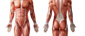
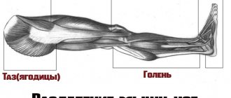
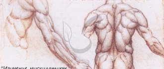
Features of the anatomy of the femoral biceps
The semimembranosus, semitendinosus muscles and biceps femoris are located on the posterior surface of the limb, and are conventionally combined into one group, called the hamstring group in English literature.
The biceps is located on the outer edge of the back of the thigh. The muscle got its name due to the presence of two heads - short and long. With a long head, it is attached to the ischial tuberosity of the pelvic bone with the help of a short flat tendon, the short head is located under the long one and is attached to the lateral surface in the lower third of the femur. At the long head is the superior bursa of the biceps femoris muscle.
Both heads at the junction form a wide belly, which below turns into a long narrow tendon, part of the bundles of which is attached to the fibula, and the other part is woven into the fascia of the leg. Between the collateral ligament and the tendon in the lower part is the lower muscle bursa, called the subtendinous.
Medial group
The pectineus muscle (m. pectineus) (Fig. 90, 129, 130, 132) flexes and adducts the thigh, rotating it outward. The flat muscle is quadrangular in shape, originates on the crest and superior ramus of the pubis, and is inserted on the medial lip of the linea aspera of the femur below the lesser trochanter.
The thin muscle (m. gracilis) (Fig. 90, 129, 130, 132, 134, 145) adducts the thigh and takes part in flexing the tibia, turning the leg inward. The long, flat muscle is located just under the skin. Its point of origin is on the lower branch of the pubic bone, and its attachment point is on the tuberosity of the tibia. The gracilis tendon fuses with the sartorius and semitendinosus tendons and the fascia of the leg to form the superficial pes anserine. The so-called goose bursa (bursa anserina) is also located here.
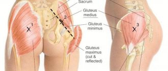
| Rice. 133. Muscles of the pelvis and thigh, side view 1 - latissimus dorsi; 2 - external oblique abdominal muscle; 3 - gluteus medius muscle; 4 - gluteus maximus muscle; 5 - sartorius muscle; 6 - muscle that tightens the fascia lata of the thigh; 7 - iliotibial tract; 8 - the longest rectus femoris muscle; 9 - biceps femoris muscle: a) long head, b) short head; 10 - vastus lateralis muscle; 11 - calf muscle |
| Rice. 134. Muscles of the pelvis and thigh, rear view 1 - gluteus maximus; 2 - adductor magnus; 3 - iliotibial tract; 4 - tendon jumper of the semitendinosus muscle; 5 - semitendinosus muscle; 6 - biceps femoris muscle; 7 - thin muscle; 8 - semimembranosus muscle; 9 - sartorius muscle; 10 - plantaris muscle; 11 - gastrocnemius muscle a) medial head, b) lateral head |
The long adductor muscle (m. adductor longus) (Fig. 90, 129, 130, 132) adducts the thigh and takes part in its flexion and outward rotation. This is a flat muscle, shaped like an irregular triangle and located on the anteromedial surface of the thigh. It starts from the superior ramus of the pubis and inserts on the middle third of the medial lip of the linea aspera of the femur.
The short adductor muscle (m. adductor brevis) (Fig. 131) adducts the thigh and takes part in its flexion and outward rotation. It is a triangular-shaped muscle that originates on the anterior surface of the inferior ramus of the pubis, lateral to the gracilis muscle, and is inserted on the upper third of the medial lip of the linea aspera of the femur.
The large adductor muscle (m. adductor magnus) (Fig. 129, 130, 131, 132, 134) adducts the thigh, partly rotating it outward. Thick, wide, the most powerful muscle of this group, located deeper than the other adductor muscles. Its point of origin is located on the ischial tuberosity, as well as on the branch of the ischium and the lower branch of the pubic bone. The attachment site is located on the medial lip of the linea aspera and the medial epicondyle of the femur. Several holes are formed in the muscle bundles, allowing blood vessels to pass through. The largest of them is called the tendon opening (hiatus tendineus). Above it is a fascial plate, and between it and the muscle a triangular-shaped space is formed, called the adductor canal (canalis adductorius) (Fig. 131). The femoral vein, artery and hidden nerve of the lower limb pass through it.
Functions of the biceps muscle
Functions of the biceps femoris:
- hip extension;
- shin flexion;
- outward rotation of the tibia;
- supination of the lower leg;
- extension of the torso from a forward bending position;
- moving the leg back;
- maintaining body balance.
The biceps femoris muscle accounts for up to 60% of the force, while its antagonist, the quadriceps, only 40%. Insufficient development of the muscles of the back of the thigh in relation to the quadriceps muscle often causes improper distribution of loads during sports, and, as a result, injuries.
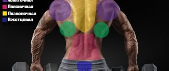
Back muscles
Mostly the flexor muscles are located here.
Double-headed
Thanks to it, we are able to rotate and maintain balance. Represented by two heads:
The first one comes from the ischial tuberosity, the second one originates from the lateral zone of the linea aspera. They are attached to the head of the fibula and are located closer to the lateral edge.
Semitendinosus
Located in the medial part. It begins in the ischial tuberosity, passes near the knee, and attaches to the tuberosity. The center is often interrupted by an oblique tendon bridge. Performs similar actions to the two-headed one.
Semimembranosus
The longest and flattest in its class. The initial end is connected to the pelvis and goes around the epicondyle.
It is interesting that it is the muscles of the posterior surface that are deprived of the proper load. Modern people have a sedentary lifestyle. This leads to less use of them, which leads to an imbalance of development and an increased risk of various injuries, especially the knee. And they are extremely unpleasant, as they limit movement and take an extremely long time to heal. Re-injury is also possible, which will lead to a longer recovery period.
Biceps injuries and their causes
Typical injuries are sprains and torn ligaments. In rare cases, the biceps femoris may be torn from its attachment site. Injuries most often occur as a result of:
- excessive stress during sports;
- uneven distribution of loads between the biceps and quadriceps muscles of the thigh;
- sudden movements and jerks;
- performing physical activity without preliminary warm-up;
- blows, bruises, twisted legs, falling due to slipping;
- general muscle weakness.
The main symptoms of injury are:
- sharp pain that occurs when moving;
- a characteristic click that occurs when a ligament ruptures;
- swelling and redness of tissue in the affected area;
- formation of subcutaneous hematomas at the site of muscle fiber damage;
- limited mobility of the limb, or, in the case of complete separation of the ligaments, its pathological mobility;
- dysfunction of the limb, inability to walk with support on the injured leg.
What to do if your quadriceps muscle hurts?
The function of the quadriceps muscle is to extend the leg at the knee joint. Stress points (SP) appear in this area due to injuries of various origins. The appearance of the above points can provoke excess tension during prolonged squatting, bending the knees, kicking, or intense cardio exercise. Hyperactivity of the 4th chapter muscle tissue provokes the occurrence of TN. This happens when skiing, dancing and walking long distances. Such points often occur in football players, basketball players, climbers, cyclists, and the like.
Any active activity that involves frequent bending and straightening of the legs can cause problems. TN in the area of muscle tissue are the main source of pain in the knee joint.
The vast medial muscle tissue places the TN in the middle of the thigh (inner surface). They provoke pain in the front and back of the knee joint and in the lower thigh area. In some cases, the pain continues for several weeks and then suddenly disappears. Weakness occurs in the knees, and they involuntarily bend.
Important! Use your thumb to locate stress points in this area. If pain is detected, you should press on these points.
A large number of TNs may appear in the vastus lateralis muscle tissue. They are located along the entire length of the muscle tissue. In most cases, they bring pain on the outer thigh. In some cases, a person is bothered by painful sensations in the “lying on his side” position.
Due to intense contractions of the wide lateral muscle tissue, the level of mobility of the knee joint may decrease, which will cause pain and difficulty walking. In some cases, a person experiences difficulty bending his leg.
Treatment methods for biceps femoris injuries
Therapy depends on the severity of the injury: for tears and sprains, treatment is conservative; with complete separation of the ligaments from the attachment site, the only treatment option is surgery to restore tissue integrity.
Conservative treatment of injuries is carried out comprehensively and includes:
- immobilization of the affected limb with a tight bandage;
- taking painkillers, injection blockades to relieve pain;
- taking non-steroidal anti-inflammatory drugs;
- cold compresses to reduce tissue swelling.
At the rehabilitation stage and in the postoperative period, physiotherapeutic procedures are prescribed to accelerate tissue regeneration and restore limb function:
- UHF heating;
- magnetic therapy;
- paraffin therapy;
- laser therapy;
- massage;
- balneotherapy;
- physiotherapy.
Exercises for hamstrings and features of the training process
The uniqueness of the biceps femoris muscle lies in its ability to shorten, so it is necessary to perform exercises in isolation aimed specifically at training them.
Before performing the exercises, it is necessary to perform a joint warm-up, and then a warm-up set of exercises, which can be replaced by walking at a calm pace with a slight incline. All exercises should be performed smoothly, without making sudden jerks or accelerations. The training complex should be completed with stretching exercises to maintain muscle elasticity.
Weighted squats
Initially, you should work with your own weight, then gradually move on to weighted squats
Depending on the level of training and physical fitness of the person performing the exercise, dumbbells or a barbell can be used as a weighting agent. The technique of squats will be the same in any case, the only difference is that when squatting, the barbell is placed on the shoulders, and the dumbbells are held with arms extended forward.
- The legs are spaced 50-60 cm wide, the toes are slightly turned outward.
- Squatting from the starting stance is performed with a straight back, so that the knees do not cross the visual line of the toes. In other words, the movement should resemble sitting on a chair, while the pelvis is pulled back.
- When squatting, the legs are bent at the knees to a right angle, so that the thighs are parallel to the floor. It is not recommended to squat lower, so as not to create excessive stress on the muscles and ligaments.
Romanian deadlift
The Romanian, or deadlift, is a basic exercise with a barbell and allows you to work out the biceps femoris muscle well. The exercise requires precise execution, preferably under the supervision of a qualified trainer.
Romanian deadlift is performed with a straight back
If the exercise is performed incorrectly, it creates excessive stress on the spinal column, which can lead to displacement of the intervertebral discs.
- When doing this, you must keep your back straight. Starting position – legs are placed at a distance slightly less than shoulder width apart, parallel to each other, the barbell is in straight arms lowered down.
- The tilt should begin by moving the pelvis back. When tilting, the bar of the bar should be as close as possible to the legs and slide along them.
- At the lowest point, you should never round your back. As you exhale, you return to the starting position, straining the back of your thighs.
- All movements should be made smoothly, without sudden jerks or accelerations.
Hyperextension
Classic hyperextension on a machine helps strengthen the muscles of the back and lower back, but if the exercise is slightly modified, it will help to isolated the muscles of the back of the thigh, in particular the biceps muscle. In order for the main load to fall on the thigh muscles, the emphasis should be placed not in the upper part of the pelvis, but at the beginning of the quadriceps, in the upper part of the thigh. It is very important to perform the exercise with a straight back to relieve the load on the lower back. Hands can be crossed on the chest or clasped at the back of the head.
Regularly performing exercises on the back of the thigh helps strengthen the biceps muscle, making the ligaments stronger and more elastic. Compliance with the correct technique during training will not only reduce the likelihood of injuries to muscle and ligamentous tissue, but will also serve to prevent similar injuries in everyday life and other types of physical activity.
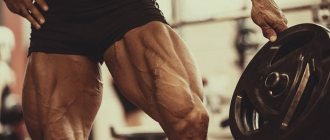
What is it - quadriceps femoris, anatomy
The concept includes 4 different elements, evenly spaced relative to each other. Due to their location and characteristics of use, the component units received their names: medial, lateral, straight and intermediate.
This is the strongest muscle complex. Therefore, for the most part, the distribution of forces falls on him. And where the pressure is greater, there is a higher risk of rupture or stretching. At a minimum, walking is responsible for the use of force. After all, in this way the human body is held vertically. In fact, the hero of the review bears all your weight when moving.
A common question is: what is it – the quadriceps, where is this object located. It is noteworthy that this is the zone under discussion - the Black Sea Security Zone. Square is four, so it’s not difficult to guess.
Start
This category includes all four main parts. In anatomy, they are often divided into three sections, the lateral line, the medallion lip, and the intermediate line. The direct area refers to the last section.
Attachment
One side is the tibia, to which all the components are attached with the help of ligaments. The second is the knee. Fixation specifically occurs at the patella. Through the same connections.
Innervation
Every tissue in the body has its own nerve endings. This is how they become one with the human nervous system. Thanks to this, we can feel touch and feel pain. Accordingly, the process of innervation is the filling with nerve endings. And if we talk about CMB, then the main function is performed by the femoral nerve of the lumbar plexus. Next, we will specifically go through the muscles under discussion.
Straight
In the structure of the quadriceps, this is the main part. It is the front, and in addition, the longest of all four. Consists of two heads, one curved, the other flat. In its foundation, in the upper half, lies a thin tendon, and the base is another – a narrow one. Which, in turn, goes into the general group of tendons.
Medial
This is a muscle already located below. It starts from the medial lip, in the area that is officially called rough. It ends on the wide tendon, where it unites with the straight line. And just below it is also attached to the patella.
Lateral
Almost the entire lateral area is given over to it. It is quite large, slightly combined, overlapped by others. It is attached to the patella in the lateral region and, like the previous ones, passes at the end into the common tendon.
Intermediate
This is the shortest and also the weakest of the groups described above. It is located between the medial and lateral, covered in front by the straight line. It passes into the tendon earlier than others, almost half as long as others due to this factor.
Having examined the description and photo of the anatomy of the 4 chapters of the thigh muscle, it’s time to move on to the tasks that they perform in the body.