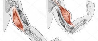
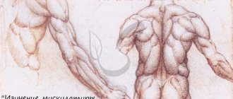
Forearm
The forearm is located between the elbow and wrist joints. Its functions include: flexion of the fingers and wrist, compression of the palm, and flexion of the arm at the elbow joint. All muscles of the forearm can be divided into 3 groups:
- Front
- Lateral (Radial)
- Rear
They are separated from each other by fascial partitions. Fascia is a kind of muscle sheath. Also, the anterior and posterior muscle groups are located in several layers.
Arm muscles
Front at four:
- Superficial. These include muscles such as: Pronator teres, Flexor carpi radialis, Palmaris longus and Flexor carpi ulnaris.
- Subsurface. These include the superficial flexor digitorum.
- Deep first layer. These are the muscles located under the superficial, but not the deepest. These include two muscles: the flexor pollicis longus and the flexor digitorum profundus.
- Deep muscles. These are the muscles that lie deepest. Also one muscle is the Pronator Profundus.
Back at two:
- Superficial. It includes muscles such as: Extensor carpi ulnaris, Extensor digitorum, and Extensor digitorum.
- Deep. It contains: Supinator, Abductor pollicis, Extensor pollicis brevis, Extensor pollicis longus and Extensor pollicis.
As we said earlier, at the initial stage when you just come to the gym, you should not devote a lot of time to training your forearms. They are already well involved during basic exercises. Especially when you have to hold heavy weight. Then, over time, you can perform 1-2 special exercises for this muscle group.
Knowledge is the root of success
Knowledge of human anatomy will certainly help an athlete consciously achieve planned results. And work specifically on muscle configuration. Here we must remember that body architecture should be studied in a complex manner, evenly paying attention to all muscles. Then you will achieve a harmonious texture.
Therefore, bodybuilders have not only the goal of their work in the gym, but also a consistent pattern of exercises, which muscles and how to pump them up, and with what tools. Muscle complexes for individual training are outlined by training days, since we all differ in our constitution.
If we look at the muscles of the right and left arms, so to speak, in cross-section, we will see bundle-shaped fibers that are divided into:
- anterior, superficial, and
- rear, deep.
Flexor and extensor muscles of the shoulder and forearm. Each has its own names. Those responsible for flexion movements are called:
- shoulder;
- two-headed;
- coracobrachial.
And the second row of responsibility is for the extensor muscles -
- ulna;
- triceps.
Most of all we talk about biceps, triceps, deltoids. These are the superficial muscles of the human arm.
The biceps is a biceps muscle, one head is long and the other is short. Thanks to it, the upper part of the arm bends, the palm and shoulder rotate.
The three-headed muscle, called the triceps, starts from the back, or rather from the shoulder blade, and extends to the elbow. With the help of this muscle, the arm bends and extends at the elbow joint and shoulder.
The forearm is represented by the following muscles:
- brachialis;
- coracoid;
- flexor carpi radialis;
- brachyradialis.
They also relate to the elbow, forearm rotation, finger extension, and drawing the arms toward the chest. By imagining how these muscles work, you understand more clearly what equipment and exercises you should choose to strengthen your arms.
Anterior, superficial muscles
Pronator teres
It is a thick muscle that has two heads:
- Large shoulder. It begins from the medial epicondyle of the humerus.
- Short elbow. It originates from the medial edge of the ulnar tuberosity.
Below, these heads unite into one muscle and are attached to the middle third of the radius. That is, this muscle connects the elbow, shoulder and forearm. This muscle is the shortest of the superficial ones.
Arm muscles
Function: Pronates the forearm. That is, it turns it clockwise in the direction of the thumb.
Flexor carpi radialis
This is a flat long muscle. It is located lateral to all the flexor muscles of the forearm. Starts from the medial epicondyle of the humerus. Next it goes down, passing to the flexor of the hand to the base of the palm and attaches to the metacarpal bone of the index finger.
Function : Flexes the wrist. Moves the hand towards the thumb. And participates in the initial phase of pronation. That is, it helps to start self-propulsion.
Palmaris longus muscle
The muscle is shaped like a spindle. Has a short abdomen. It is attached to the medial epicondyle of the humerus. Next it goes to the palm and turns into a powerful, wide tendon (aponeurosis).
Function: Flexes the hand and also stretches the tendon aponeurosis (the wide muscle fascia of a triangular shape). It is compacted by tendons. See the picture above.
Flexor carpi ulnaris
Located on the medial edge of the forearm. The muscle has a long belly and thick and strong tendons.
Two heads can be distinguished:
- Shoulder. Starts from the medial epicondyle of the humerus.
- Elbow. Starts from the olecranon and the upper ⅔ of the ulna.
Passing down, they unite into one muscle and attach to the uncinate process of the wrist and the 5th metacarpal bone.
Function: Bringing the wrist towards the body, towards the little finger. Helps flex your wrist.
These are all superficial muscles. As far as can be seen, they all perform almost the same functions, and are located on the medial side of the forearm. In order to develop them, you need to do exercises in which flexion occurs at the wrist.
Types of diseases
The forearm is located in a place where birth defects are rare.
Such vices include:
- Peromelia. In this case, the entire arm is missing, and a small rudiment may extend from the body.
- Congenital club hand. At birth, the fetus has completely absent or underdeveloped radius or ulna bones.
- Phocomelia or underdevelopment of the shoulder and forearm. It consists in the fact that a person’s hand starts directly from the body.
- Hemimelia. This defect is characterized by the complete absence of the forearm.
Frequently occurring injuries in the forearm area are classified as injuries. They are classified as closed and open injuries.
Closed injuries:
- Bruises.
- Fractures.
- Muscle or tendon ruptures.
- Compression of blood vessels, nerves, tightening of muscles, as a result of bruises, accidents.
Open injuries:
- Bone fractures without displacement.
- Extensive wounds.
- Comminuted, comminuted fractures.
Human limbs are often affected by various diseases, which are based on inflammatory processes, less often infectious causes.
These include:
- Myositis. Inflammation of one or more muscles develops against the background of overstrain and hypothermia.
- Tunnel syndrome. A neurological disease caused by compression or pinched nerves.
- Aseptic tendovaginitis. Develops after heavy physical labor, when the muscles of the forearm were involved. Most often, the lesion affects the superficial muscles located in the back of the forearm.
- Acute nonspecific tendovaginitis. It is observed in the tendon sheaths of the flexors of the fingers, located in the deep muscles of the anterior part of the forearm. The cause of development is osteomyelitis, panaritium
- Osteomyelitis. The disease develops as hematogenous, affecting all bones and surrounding tissues. Other causes of the disease: postoperative or post-traumatic (fractures, injuries, wounds).
- Tuberculosis. The extrapulmonary form of the disease affects some parts of the bones of the forearm.
- Mycoses. Diseases caused by parasitic fungi are rare, but still affect the bones of the forearm.
- Contractures of the hand and fingers. They develop as a consequence of diseases of muscles and tendons, after injuries.
- Tumors. Benign formations such as angioma, lipoma, fibroma can develop in soft tissues. Tissues also serve as a source of development of malignant tumors. Chondrosarcoma and fibrosarcoma are found in the forearm area. Metastases in this area are observed in rare cases.
Subsurface muscles
Flexor digitorum superficialis
This muscle is covered by the palmaris muscle and the flexor digitorum. It has two heads:
- Shoulder. It is a long and narrow head that originates from the medial epicondyle of the humerus. And also, from the external process of the ulna
- Radial. This head is short, but at the same time very wide. Attached to the inner part of the radius bone.
Both heads unite into a common muscle and form a large abdomen. Which ends in a tendon. And it is attached to the base of the middle phalanges except the large one.
Function: Helps to bend the hand. Bends the middle phalanges of the fingers from the 2nd to the 5th, and with them the fingers themselves.
Structural features
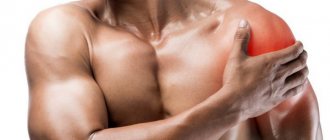
If we consider it as a complex conglomerate, the shoulder joint is formed by bones, cartilage, joint capsule, bursae, muscles and ligaments. In its structure, it is a simple, complex spherical joint consisting of 2 bones. The components that form it have different structures and functions, but are in strict interaction designed to protect the joint from injury and ensure its mobility.
Shoulder joint components:
- spatula
- brachial bone
- labrum
- joint capsule
- bursae
- muscles, including the rotator cuff
- ligaments
The shoulder joint is formed by the scapula and humerus, enclosed in a joint capsule.
The rounded head of the humerus is in contact with the fairly flat articular bed of the scapula. In this case, the scapula remains practically motionless and the movement of the hand occurs due to the displacement of the head relative to the articular bed. Moreover, the diameter of the head is 3 times larger than the diameter of the bed.
This discrepancy between shape and size provides a wide range of movements, and the stability of the articulation is achieved through the muscular corset and ligamentous apparatus. The strength of the articulation is also given by the articular lip located in the scapular cavity - cartilage, the curved edges of which extend beyond the bed and cover the head of the humerus, and the elastic rotator cuff surrounding it.
Deep muscles first layer
Flexor pollicis longus
A very long and flat muscle. Starts from the lower middle part of the radius and the edge of the medial epicondyle of the shoulder. Passes through the entire forearm and is attached to the upper phalanx of the thumb.
Function : Flexes the thumb. Together with other muscles, it takes part in flexing the wrist.
Flexor digitorum profundus
This is one of the strongest muscles of the forearm. Has a wide belly. Starts from the upper surface of the ulna. Next, the muscle fibers are directed downwards and diverge into 4 tendons. They lie in the carpal canal, under the tendons of the superficial flexor of the fingers, that is, they seem to cover them. And then, the deep flexor tendons are attached to the upper phalanges of the 4 fingers from the index to the little finger.
Function : Flexes the upper phalanges of the fingers from the 2nd to the 5th. Participates in flexion of the hand.
BICEPS
The biceps consists of two heads:
- The longus (long tendon, but the muscle is small) is located on the outer part of the arm.
- The brevis (short tendon, but large muscle) is located on the inside of the arm.
Both heads originate on the shoulder blade, just in different places... in other words, both heads are connected into one tendon, which is located next to the elbow joint. Subsequently, both heads form a common belly, which passes into a powerful tendon (the tendon itself is attached slightly inward (to the side of the forearm)), which is attached to the radius, and despite their name, both heads have the same length, because the long head has in fact, a longer tendon with which it is attached below to the bone.
The biceps flexes the forearm and rotates it outward (this is supination), which means that in addition to the fact that the biceps can simply bend the arm at the elbow joint, it can also supinate it (i.e., turn the palm towards the thumb).
Through the short head, the biceps takes part in adducting the arm, and through the long head, abducting the arm.
In addition to the biceps, the anterior group of shoulder muscles also consists of the brachialis muscle, which is located below the biceps, as if pushing it out. The main function is to flex the forearm.
FOCUS ON THE BICEPS HEADS
According to statistics, there are no problems with the development of the short head (the one located on the inside of the arm), it responds perfectly to the load, and grows well from any bending of the arm. But with the development of the long head, the one that is located on the outer part of the arm, most people have problems!
Treatment
- In order to fight the outer head (long), you need to move your elbows as far behind your back as possible, this is the only way to engage the outer part of the biceps.
- In order to fight the internal head (short), on the contrary, you need to bring your elbows forward as much as possible.
GRIP when working on BICEPS
- The wider your grip, the more the inner head will work.
- The narrower your grip, the more the outside head will work.
In general, I don’t recommend a narrow grip. In theory, the narrower the grip, the more you will bring your elbows forward, and based on the above (if the elbows are brought forward), then the inner head is strongly activated.
Lateral (radial) group
This includes the muscles of the front and back of the forearm, which are located on its lateral part, that is, the outside. These include: brachioradialis, extensor carpi radialis longus, extensor carpi radialis brevis.
Brachioradialis muscle
A large muscle located on the lateral side of the forearm. It is attached from above to the lateral edge of the humerus, slightly above the lateral epicondyle. It runs along the forearm and attaches to the very bottom of the radius.
Function: Bends the arm at the elbow joint. Directly involved in pronation and supination of the radius.
Extensor carpi longus
From the name it is clear that the muscle itself is very long. It has a spindle-shaped shape. It begins with a narrow tendon from the lateral epicondyle of the humerus, and is attached to the back of the metacarpal bone of the index finger (the bones that stick out when the palm is clenched into a fist).
Function : Extends the hand and moves it towards the thumb.
Extensor carpi radialis brevis
This muscle is not large, covered by a long extensor muscle. It starts from the lateral epicondyle, goes down and passes into the tendon. And attached to the back of the metacarpal bone of the middle finger.
Function: Extension of the hand and abduction towards the thumb.
Works in tandem with the previous muscle.
Shoulder joint: muscles, ligaments, bones
Without muscle tissue, ligaments, and bones, movement is impossible. Only thanks to the integrated functioning of these components does the joint work.
Muscle work in the shoulder joint
Thanks to muscle tissue, ligaments, and nerve impulses, flexion, extension and other actions occur in the articular surface. Different types of muscles are responsible for different movements. They fix the bone components, and ligaments and nerve endings bring them into a certain position.
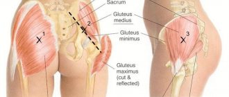
The figure below lists the muscle groups responsible for one or another part of the shoulder joint.
There are the following types of muscles in the shoulder joint:
- deltoid
- supraspinatus
- infraspinatus
- coracobrachial
- chest
- subscapular.
The deltoid, chest muscles, and coracobrachial muscle tissue are responsible for bending the arm
The deltoid muscle, a large rounded, infraspinatus muscle tissue, is also responsible for extending To move your arm to a horizontal position , you will have to use the supraspinatus, deltoid muscle. And in order to raise the arm higher they use a trapezoidal, diamond-shaped, or large round one.
Internal turns are carried out by the anterior bundles of the deltoid muscle, pectoralis major, orbicularis, and subscapularis muscle tissue. External turns are made by the posterior bundles of deltoid muscle fibers, teres minor, and infraspinatus.
The structure of the muscle tissue of the shoulders and neck
Blood flow of muscle tissue
In this zone there is an axillary artery, which then flows into the brachial artery. Due to this blood vessel, oxygen, glucose and other substances are delivered to the joint. When all components, from veins to connective fibers, work harmoniously, there will be no problems with the functioning of the joint.
All muscle tissues have a blood supply, which is carried out by vessels. Thus, the thoracic arteries provide blood flow to the thoracoacromial, lateral, intercostal anterior artery, and intercostal posterior artery. They are controlled by pectoral nerve endings, lateral nerves and medial nerves.
Ligaments - their role
You can see the ligaments of the articular surface of the shoulder in the image below; as you can see, there are many such fibers, and they are all responsible for a certain area of the shoulder. And they are the ones that set the joints in motion.
There is also a cavity that reduces joint friction thanks to synovial fluid. This fluid is found between the cartilage and bones of the joint. This is precisely why a person’s movements are smooth and there is no friction or pain.
So, the shoulder joint includes the following ligaments:
- Acromioclavicular.
- The coracoclavicular ligament connects the corresponding bony surfaces (see picture below).
- Coracoacromial.
- Articular-brachial ligaments of the upper and middle part of the shoulder.
- Inferior glenohumeral ligament.
Ligaments
The shoulder joint is the most complex in terms of mobility. Because other joints do not have as much ligamentous tissue as here. In order for the ligaments and the joint itself to work well, in any case, muscle tissue is also necessary, which prevents the shoulder joint from dislocations and the ligaments from sprains. It is also important that the synovial bursa retains fluid. The capsule will be in perfect order if strong ligaments maintain it. These include the coracobrachial and articular-brachial ligaments.
Bones and cartilage
Cartilage tissue is not nourished by the capillary network; in particular, hyaline tissue does not contain capillaries, and therefore does not supply oxygen and other necessary components through the bloodstream. This action is provided by synovial fluid, in direct contact with cartilage tissue by the diffusion method. In order for the cartilage tissue to remain intact, it is necessary that the liquid does not change its quality and that it comes in the right quantity. Otherwise, the condition of the cartilaginous surfaces, and then the joint itself, will be disrupted.
The bone of the shoulder joint itself looks like a ball-and-socket joint. On top of the humeral bone itself there is a spherical head. You can see the shoulder blade nearby; it goes into the waist part at the top. It has the same spherical depression as the head of the humerus. It just fits into the depression, but only partially. Its size is almost four times smaller than this head.
Bone joint
Those bones mentioned above are the parts that form the joint. Its structure is such that even when a person raises his arms up or makes other movements with them thanks to muscles, nerve impulses, ligaments, the head of the shoulder is held opposite the socket of the scapula.
As you can see, the mechanism is not so difficult to understand, the main thing is to study all the components - muscle tissue, bone surfaces, ligaments and other elements of the shoulder joint. Then it will become clear why you can make any movements with your hands thanks to this mechanism.
Posterior superficial muscles
Extensor carpi ulnaris
This muscle is located along the inner edge of the back surface of the forearm. It has a spindle-shaped long abdomen. Starts with two heads:
- Shoulder. It originates from the lateral epicondyle of the humerus, slightly above the insertion of the extensor of the little finger.
- Elbow. Starts from the posterior edge of the ulna, at the level of the lower part of the olecranon muscle.
Both heads connect and pass into a short but very powerful tendon. And it is already attached to the posterior surface of the 5th metacarpal bone. That is, to the little finger.
Function: Extends the hand. And he takes it to the side of the elbow.
Extensor digitorum
Just like the previous muscle, it has a spindle-shaped abdomen. It originates from the lateral epicondyle of the humerus, also the articular capsule of the elbow joint and the fascia of the forearm. It goes down and closer to the wrist turns into 4 thin tendons. They pass under the extensor retinaculum. Having passed to the hand, the tendons are connected to each other by permanent, thin intertendinous connections. They go to the base of the lower phalanges of the fingers, from the index to the little finger and end there with tendon stretches. Next, the tendon is divided into three legs, of which 2 lateral ones are attached to the base of the upper phalanges of the 4 fingers. And the middle leg to the middle phalanx.
Function : Extends fingers. Participates in the extension of the hand and abduction of the fingers from each other.
Extensor of the little finger
It is a small abdomen, spindle-shaped. The muscle begins from the lateral epicondyle. It goes down and passes into the tendon, which connects to the extensor tendon of the fingers going to the little finger. And attached together with them to the upper phalanx of the 5th finger.
Function: Extends the little finger.
Deep muscles
Arch support
It has the shape of a rhombus. This is a thin muscle plate located on the back of the forearm. Attached from above to the lateral epicondyle, as well as to the crest of the supinator of the ulna and the capsule of the elbow joint. It is directed obliquely downwards and is attached to the radius below its tubercle.
Function: Pronates or supinates the forearm.
Abductor pollicis longus muscle
It has a two-fingered abdomen (that is, they look like feathers) turning into a thin tendon. It starts from the posterior surface of the radius and ulna. Heading down the forearm, bending around the radius, and attaches to the posterior base of the 1st metacarpal bone. That is, to the thumb.
Function: Moves the thumb to the side.
Extensor pollicis brevis
This muscle is located in the lower back surface of the forearm. It starts from the interosseous membrane and the dorsum of the radius. It goes obliquely down and is located next to the tendon of the long muscle, and is attached to the base of the posterior surface of the middle phalanx of the thumb.
Function: Extends the thumb. It also moves the middle phalanx a little to the side.
Extensor pollicis longus
This muscle has a fusiform belly and a long tendon. Located next to the short extensor muscle. It originates from the interosseous septum of the posterior surface of the ulna. It goes down and goes into the tendon. Which is attached to the back surface of the upper phalanx of the thumb.
Function: Extends the thumb. And also takes it a little to the side.
Extensor index finger
The muscle is located in the lower half of the forearm. It has a narrow and long abdomen. Starts from the lower third of the posterior surface of the ulna. At the bottom it passes into the tendon, and is attached to the back of the upper phalanx of the index finger. Sometimes this muscle is absent.
Function: Extends the index finger.
These are all the available muscles of the forearm. As far as you can see, they are responsible for important functions, helping to bend and straighten your fingers. And also, they move the wrist. During basic exercises, they act as stabilizer muscles. Which help maintain weight. Their good development will help you work without additional equipment (straps, wristbands). It also protects the wrist from all kinds of injuries and sprains.
Abductor pollicis longus (musculus abductor policis longus)
The supinator is the most “radial” muscle of all the muscles in this layer. Now we begin to move from it in the ulnar direction, and the first thing we notice is a rather long, triangular-shaped muscle that goes towards the big toe. We are talking about the abductor pollicis longus muscle.
With a thin green line, I also highlighted the tendon that attaches the muscle to the tendon extension of the thumb.
Location: Starts from the posterior surface of the radius and ulna, attaches to the base of the first metacarpal bone (that is, the one in front of the phalanges of the thumb).
Function: abduction of the thumb.
Hand bones
The hand consists of the bones of the wrist, as well as the bones of the fingers (phalanx) and metacarpus (tubular bones running from the wrist to the fingers). The wrist itself has eight spongy bones. They are short in size and arranged in 2 rows:
- Upper. It includes such bones as: scaphoid, triquetrum, pisiform and lunate.
- Lower. Consists of: trapezoid, capitate, trapezoid and hamate.
The bottom row connects to the top and the carpal bones. And also among themselves. And they form a low-moving joint.
After the carpus come the metacarpal bones. There are only five of them, one for each finger. Next they connect to the phalanges of the fingers. They have a short tubular shape. Each finger has three phalanges: main (proximal or lower), middle and upper (distal or terminal). The exception is the thumb; it consists of only two phalanges - lower and upper.
Muscles of the hand
The hand can be divided into 3 groups, which contain several muscles:
Muscles of the thumb
It includes the following muscles:
- Abductor pollicis brevis muscle . It is located just under the skin, on the side of the thumb. It starts from the tubercle of the scaphoid, fascia of the forearm and tendons of the long muscle. And is attached to the lower phalanx of the thumb.
- Flexor pollicis brevis. It is also located immediately under the skin, next to the short muscle, closer to the inside of the palm. It starts from the trapezoid bone, trapezoid, capitate bone and the base of the 1st metacarpal bone. Divides into 2 heads: the superficial one is attached to the sesamoid bone, the outer part. And the deep one is attached to the metacarpal phalanx of the thumb.
- Contrasting . A thin, triangular-shaped muscle. Located under the short muscle. Originates from the tubercle of the trapezium bone. And is attached to the outer edge of the metacarpal bone of the thumb.
- Adductor pollicis. It has the deepest location of the muscles of this group. It has two heads that point towards each other. Bony: starts from the capitate bone and 2-3 metacarpal bones. Transverse: from the outer surface of the 3rd metacarpal bone and their heads 2 and 3 bones. then they converge at an angle and attach to the base of the middle phalanx of the thumb,
Function: From the name you can understand what function these muscles perform. They flex, abduct, oppose the little finger (bring them together) and adduct the thumb.
Muscles of the little finger
It includes:
- Palmaris brevis muscle . This is a thin muscle plate with parallel bundles. It starts from the inner edge of the palmar aponeurosis. And the lower edge is woven into the skin of the eminence of the little finger.
- Abductor of the little finger . It is located on the far edge of the palm, immediately under the skin. Originates from the flexor carpi tendon, pisiform bone, and flexor retinaculum. The lower edge is attached to the medial edge of the middle phalanx of the little finger.
- Opposite muscle . Located lateral to the previous muscle. Originates from the hamate and the flexor retinaculum. And attached to the ulnar edge of the metacarpal bone of the little finger.
- Flexor pollicis brevis . Slightly covered by the palmaris brevis muscle and skin. The upper edge is attached to the hamate bone, and the lower edge is attached to the ulnar edge of the middle phalanx of the little finger.
Function: They perform the same functions as the thumb muscles. Bend and retract the little finger. They also bring it to the thumb.
Middle group
Comprises:
- Vermiform muscles . These are 4 spindle-shaped muscles. They are attached to the radial part of the tendon of the deep flexor of the digitorum. And they are attached to the back surface of the lower phalanx of the 4 fingers. From the index finger to the little finger.
- Palmar interosseous muscle . These are the three interosseous muscles. Which are attached to the ulnar side of the 2nd metacarpal and radial 4th and 5th bones. Having passed into the tendon, it is attached to the lower phalanges of the 2nd, 4th and 5th fingers.
- Dorsal interosseous muscles . Located on the back surface of the metacarpus. These are 4 muscles with two so-called muscle feathers. Each muscle has two heads, which are attached to the base of two adjacent metacarpal bones. Next, the 1st and 2nd are attached to the radial edge of the middle and index fingers, and the 3rd and 4th are attached to the ulnar edge of the middle and ring fingers.
Function: Flexion of the lower phalanx of 4 fingers. The palmar muscles bring the fingers to the middle, and the back muscles move them apart.
As far as you can see, the forearm and hand muscles are interconnected, which is why they train together.
I suggest watching a 5 minute video. From it you can find out the top 5 best exercises for training your forearms and hands.
Anatomy of arm muscles: how to train correctly
Let's go over the anatomical features of the arm muscles and, as a result, derive some rules for their effective training. And we'll start with...
No. 1. Biceps
The biceps is a superficial muscle, so the indicative appearance of your hand muscles will depend on its quality development. The main movements in which he participates are lifting the projectile from bottom to top, i.e. bringing it to the chest. To create the peak of the biceps, it is necessary to use lifts with supination during the exercise - turning the hand upward when the palm faces the ceiling and the little finger is located above the thumb, or lifts with an already supinated hand.
The best exercises for biceps:
- standing barbell/dumbbell lifts (straight/EZ bar);
- reverse grip pull-ups;
- dumbbell lifts while sitting at an upward angle from an extended position;
It is worth understanding that the shape of the biceps is laid in you by Mother Nature; it can be long with short ligaments or short with long ends of the ligaments (like Schwarzenegger).
No. 2. Triceps
The triceps make up 2/3 of the volume of the arm, therefore, if the arms do not have enough volume, then it is necessary to first “hammer” the triceps and only then the biceps. The main “profession” of all three triceps heads is extension of the arm at the elbow joint, while the medial one is the most active of all heads. Triceps antagonists (biceps, brachialis) are physiologically more powerful than the triceps muscle, which is manifested in a slight bend of the arms at the elbow when they hang freely during rest.
For the qualitative development of the triceps brachii muscle, it is necessary to use flexion/extension exercises with free weight. Quality means an increase in the volume-strength characteristics of a given muscle group. You should not devote time to isolated exercise machines (guys, leave them to the girls); it is better to use multi-joint exercises in which all 3 heads of the triceps are immediately “captured” into work.
The best triceps exercises:
- reverse push-ups from a bench;
- dips;
- close grip bench press.
No. 3. Forearm muscles
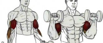
High-quality anatomy of the arm muscles requires good development of this muscle group. The brachialis muscle creates a supporting platform for the biceps, as if pushing it to the “surface”. The brachialis is activated by static elbow flexion and works in all biceps exercises, but it is best engaged during reverse-grip biceps curls.
The brachioradialis (brachyradialis) muscle is actively involved in the work when lifting dumbbells with a hammer grip, i.e. when the thumb points up. The coracoid muscle plays an important role in the development of hand muscles and is clearly visible in the double biceps pose in front. The best way to hit the coracoid muscle is to do dumbbell lifts in front of you, dumbbell flyes while lying on a bench.
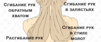
The best exercises for the forearms:
- spider curls (barbell lifts with a reverse grip);
- Hammer lifts (lifting dumbbells with a hammer grip);
- wrist straight/reverse lifts of the barbell from a bench while kneeling.
Uff-f, well, that’s all, actually, now let’s sum it up and say goodbye.