What are the muscles that act oppositely called?
Human musculature is designed in such a way that many muscles have “brothers” that perform completely opposite work: at the moment when one muscle tenses, the opposing one relaxes, and vice versa.
These muscles - flexors and extensors, which control the movement of the human body or individual limbs in space, are called antagonists. This is exactly how a person makes movements - thanks to a control system strictly coordinated by the brain and the coordinated work of the muscles that move the skeleton.
How do they work?
The brain sends an impulse to the nerve endings of a muscle, such as the biceps of the arm, and it contracts, bending the arm. The triceps - the extensor of the arm - is relaxed at this moment, since the brain gave the corresponding signal to it.
Flexor and extensor muscles, that is, antagonists, always work harmoniously, mutually replacing each other, but sometimes they can work simultaneously, maintaining a motionless, that is, static position of the body in space. A striking example of such work is the well-known plank pose, in which the body hangs motionless above the floor, resting only on the hands and toes. Most of the main flexors and extensors of the muscles in this position do exactly half of the work required for them, as a result, the body maintains this position. If a person does not strain, say, the abdominal muscle, then it becomes difficult for his back, since under the pressure of gravity the lower back begins to sag and sag. Arms lowered down along the body are completely relaxed antagonist muscles, and an outstretched arm in front of you at shoulder level is synchronous work of both muscle groups.
What determines the quality of movement?
The quality of the flexor and extensor muscles depends on several factors:
- The amplitude of movement mainly depends on the length of muscle fibers and the factors restraining them, for example, muscle spasm or post-traumatic scar greatly reduces the range of movement, and elasticity and good blood flow, on the contrary, significantly add amplitude to muscle work. That is why it is important to warm up the body well with dynamic movements before training in order to saturate the muscles with blood.
- The strength of a muscle depends on two aspects: the amount of leverage that the muscle uses, and directly the number and thickness of the muscle fibers that make it up. For example, lifting a 10 kg weight using the entire length of your arm is easy (large lever), but lifting it with just your hand will be more difficult. The same is true with the number of muscle fibers: a muscle that is 5 cm in diameter is several times stronger than one that is only 2 cm thick.
- All muscle movements are controlled by the somatic nervous system, therefore all body movements, especially the coordinated actions of the flexor and extensor muscles, depend on the speed and quality of its work.
If an athlete knows about the correct functioning of muscles, his training becomes more conscious and therefore correct, the level of efficiency increases significantly with less energy expenditure.
Examples of antagonist muscles
The simplest examples of flexor and extensor muscles:
- The femoral biceps and quadriceps are the flexor and extensor muscles of the leg, or more precisely the hip. The biceps is located at the back, attached to the ischium at the top and bottom, turning into a tendon, adjacent to the femur in the area of the knee joint. And the quadriceps is an extensor, located on the front side of the thigh, attached by a tendon to the knee joint, and the upper part is attached to the pelvic bone.
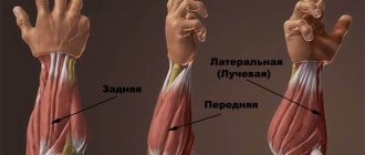
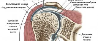
- The biceps and triceps are the flexor and extensor muscles of the arm, located between the elbow and shoulder joints and attached to them by powerful tendons. They are the main muscles that form the shoulder and control the vast majority of flexion and extension movements of the arm.
You can often notice that if there is an overly active extensor muscle, then, as a result, the flexor muscle will be in a passive state, that is, not sufficiently developed, which creates inadequate body movements with a greater loss of energy than in harmoniously trained people (yogis are an example of this) .
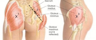
Functions of the back muscles
The fact that the human body contains such a large number of spinal muscles is associated with the differentiation of the whole body and the spine in particular. The vertical position of a person provides the power of this muscle. Without it, the body would bend forward. After all, it is in front of the spine that the center of gravity lies. In addition, this group also includes some muscles that lift the torso. Agree, their significance is very great.
The group of back muscles associated with the upper limbs is located in 2 layers. The trapezius and latissimus muscles lie in the superficial layer. The second contains the rhomboid and also the levator scapulae.
In addition to the above-described meaning, the muscles of the upper limb located on the torso have another. For example, those that attach to the shoulder blade do more than just move it. They fix the scapula when antagonistic muscle groups contract simultaneously. In addition, if a limb is immobilized by the tension of other muscles, then when they contract, they no longer act on the limb itself, but on the chest. They expand it, that is, they act as auxiliary muscles of inspiration. The body uses these muscles during difficult and increased breathing, in particular during physical work, running or respiratory diseases.
So, we looked at the main muscles of the torso. Anatomy is a science that requires deep study. A superficial consideration of individual issues does not allow us to see the entire system as a whole. Meanwhile, the muscles of the trunk and neck are only part of the complex mechanism with which we control our body.
Another example of antagonist muscles
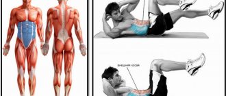
The rectus abdominis and longitudinal muscles along the spine, along with the lumbar muscle, are also prominent representatives of the flexors and extensors of the body, and they are the most global, because thanks to their coordinated and uninterrupted work, the human body takes on various positions in space: from a vertical position of the torso to bending in an arc or, on the contrary, bending back.
And if a person is working to correct posture: eliminate kyphosis, correct scoliotic curvature, or remove hyperlordosis in the lower back, he needs not only to work on the spinal extensors and lumbar muscles, but also to actively pump the abdominal muscles, in particular the longitudinal abdominal muscle.
Lecture No. 9. "Muscles of the trunk."
The torso is the part of the body without the head, neck and limbs. Muscles of the trunk include:
- backs
- breasts
- belly
They ensure the vertical position of the body, the movement of the spinal column and ribs, and form the walls of cavities.
Back muscles.
Paired, occupy the entire dorsal surface of the body. They lie in several layers (superficial and deep). Superficial muscles moved during development from the limbs to the back. The deep ones were formed from muscle anlages - myotomes. They partially preserved the segmental structure.
Surface:
- Trapezius muscle (m. trapecius) - occupies the upper part of the back, has the shape of a triangle, the bases of which are directed towards the spine. Taken together, the muscles on both sides form a trapezoid shape. It starts from the spinous processes of the thoracic vertebrae, the nuchal ligament and the occipital bone, and is attached to the acromial end of the clavicle, the acromion and the scapular spine. The upper part raises the scapula and shoulder girdle, the middle part brings the scapula closer to the spine, the lower part pulls it down.
- The latissimus dorsi muscle (m. latissmus dorsi) is flat, triangular in shape, and occupies the entire lower part of the back. It starts from the spinous processes of the 5-6 lower thoracic vertebrae, all lumbar vertebrae, from the iliac and median sacral crests and is attached to the crest of the lesser tubercle of the humerus. Pulls the shoulder towards the body, the arm back, while simultaneously turning it inward (conductor muscle).
- Small and large diamond-shaped (mm rombojdei major et minor) - lie under the trapezoid. From the spinous processes of the two lower cervical and 4 upper thoracic vertebrae and are attached to the medial edge of the scapula. Bring the shoulder blade closer to the spine and pull it up.
- The levator scapule muscle (m. levator scapule) lies above the rhomboids. It starts from the transverse processes of the four upper vertebrae and is attached to the upper corner of the scapula.
- The superior posterior serratus muscle (m. serratus posterior superior) is under the rhomboid muscles. It starts from the spinous processes of the two lower cervical and two upper thoracic vertebrae and is attached with 4 teeth to the 2-5 ribs. Raises the upper ribs and participates in the act of inhalation.
- The lower posterior serratus muscle (m. serratus posterior inferior) - lies under the latissimus muscle. It starts from the spinous processes of the 2 lower thoracic and 2 upper lumbar vertebrae and is attached with 4 teeth to the 9-12 ribs. Lowers the lower ribs and participates in the act of exhalation.
Deep back muscles.
Consists of 3 layers:
- Superficial (head, neck and lateral tract)
- Middle (medial tract)
- Deep (interspinous, intertransverse and deep suboccipital muscles)
The most developed muscles of the superficial layer, which perform the work of holding the body in an upright position. Middle layer muscles - straightening and rotating the spine. The muscles of the deep layer lie in the places between the spinous and transverse processes of the vertebrae in the most mobile areas: the cervical, lumbar and lower thoracic region.
- The splenius capitis muscle (m. splenius capitis) starts from the nuchal ligament, the spinous process of the 7th cervical and 4th upper thoracic vertebrae and is attached to the occipital bone and mastoid process.
- Belt muscle of the neck (m. splenius cervicis) - starts from the spinous processes of the 3-4 thoracic vertebrae and is attached to the transverse processes of the three upper cervical vertebrae. Both muscles extend the head and turn in the direction of contraction.
- Lateral tract (spinal erector muscle) – m. erector spinae:
- Iliocostal
- Longest
- spinous
- The medial tract is a group of transverse spinous muscles that spread obliquely upward through the vertebrae from the transverse to the spinous processes. The semispinalis - after 5-6 vertebrae, the middle multifidus - after 3-4 vertebrae, the deep rotator muscles - after 1 vertebra.
- The interspinous muscles of the neck, chest and lower back connect the spinous processes to each other, starting from the 2nd cervical vertebra. Extend the spine.
- The intertransverse muscles of the neck, chest and lower back are bundles that spread across the transverse processes of adjacent vertebrae. The body is tilted to the sides.
- Suboccipital muscles (m. suboczipitalis) - large and small posterior rectus heads and superior and inferior rectus heads) - lie between the occipital bone and 1-2 cervical vertebrae. With a unilateral contraction, the head is turned and tilted to the side, with a bilateral contraction, the head is thrown back.
Chest muscles.
Superficial ones move from the upper limbs and are attached to their bones. Deep (own) are attached to the bones of the chest.
Superficial chest muscles.
- The pectoralis major muscle (m. pectoralis major) is massive, fan-shaped, located in the upper part of the chest, limiting the axillary cavity in front. It starts from the medial half of the clavicle, sternum, cartilages of the upper 6 ribs and is attached to the crest of the greater tubercle of the humerus. Rotates inward and lowers the raised arm.
- The pectoralis minor muscle (m. pectoralis minor) is flat, triangular in shape, lies under 1. It starts from the 2-5 ribs and is attached to the coracoid process of the scapula. tilts the shoulder blade forward, lowers the shoulder girdle, raises the upper ribs, and participates in the act of inhalation.
- Subclavian muscle (m. subclaviae) - starts from the cartilage of the first rib and is attached to the lower surface of the acromial end of the clavicle. Pulls the collarbone down and forward, raises the first pair of ribs, and participates in the act of inhalation.
- The serratus anterior muscle (m. serratus anterior) is wide, quadrangular, attached to the side of the chest. It starts with teeth from the upper 8-9 ribs and is attached to the lower corner and medial edge of the scapula. Moves the shoulder blade forward, moves the arm above the horizontal line.
Deep chest muscles.
- External intercostal muscles (m. intercostalis externi) - 11 on each side, fill the spaces from the spinal column to the costal cartilages. Raise the ribs and participate in the act of inhalation.
- Internal intercostal muscles (m. intercostalis interni) - lie under 1, have the opposite direction of the bundles and fill the gaps from the sternum to the corners of the ribs. These are the innermost intercostal muscles. The ribs lower and participate in the act of exhalation.
- Subcostal muscles (mm subcostalis) - spread over 1-2 ribs, located in the posterior part of the chest. The ribs lower and participate in the act of exhalation.
- The transverse chest muscle (m. transversum toracis) is thin, flat, fusiform, adjacent to the inner surface of the anterior chest wall. It starts from the xiphoid process and the lower half of the body of the sternum and is attached to the inner surface of the cartilages of 2-6 ribs. Lowers the ribs and participates in the act of exhalation.
- The muscles that lift the ribs (mm levater costalis) are divided into short and long, located next to the thoracic spine under the extensor muscle of the trunk. They start from the transverse processes of the 7th cervical and 10-11th thoracic vertebrae and are attached to the corners of the ribs (short), pass through the rib to the next (long). Raise the ribs and participate in the act of inhalation.
Anatomically and functionally connected to the chest muscles, the thoraco-abdominal barrier - the diaphragm - is the main respiratory muscle. This is a flat striated muscle stretched along the lower opening of the chest - the partition between the chest and abdominal cavities. It has the shape of a dome, convexly facing the inside of the chest. Its muscle bundles in the center of the dome pass into fibrous tissue and form a tendon center. In the middle it has an indentation from the heart lying on it.
Diaphragm parts:
- sternal
- costal
- lumbar
There are 2 openings in the lumbar part: in the back - the aortic, containing the aorta and the thoracic lymphatic duct, in front - the esophageal, containing the esophagus, and in the tendon center on the right there is an opening of the inferior vena cava. When the diaphragm contracts, its dome flattens and lowers, the volume of the chest increases, which facilitates inhalation; when the diaphragm relaxes, its dome returns to its original position, the volume of the chest decreases, which facilitates exhalation.
The simultaneous contraction of the diaphragm with the abdominal muscles promotes the act of defecation, urination, childbirth in women, and also puts pressure on the liver, squeezing blood out of it into the inferior vena cava.
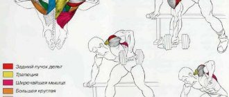
Pectoral muscles and diamond backs
These two couples also belong to the antagonists, although they are often unfairly classified in other categories. The relationship between spasm of the pectoral muscles and the passive rhomboid muscles of the back has repeatedly become an area of research for physical and yoga therapists, kinesiologists and rehabilitation specialists. The pectoralis major and minor muscles are shaped like a fan. They are located on the front of the chest, originate in one bundle at the collarbones, the lower one - at the upper abdominal wall and are attached to the crests of the humerus. Spasm of the pectoral muscles can be determined not only by the person’s stoop, but also by the position of his arms, lowered along the body. His arms from the shoulder and down to the hand will be turned inward, that is, the hands will face back with the palms.
The rhomboid muscles are located between the shoulder blades, controlling their work together with the trapezius, which, in turn, directly depend on the freedom of the shoulder muscles, in the area of which there is already an attachment of the pectoral muscles. As a result, a person works on stooping, loading the back muscles, but in fact he first needs to get rid of hypertonicity of the pectoral muscles, then work the extensor and flexor muscles of the neck, which will give freedom to his posture.
Enter site name
Additional Information
The main muscles of the chest are the pectoral muscles
, which raise the arms during contraction, and
intercostal muscles
, which raise the ribs during inhalation.
Between the chest cavity and the abdominal cavity, inside the body, there is a muscle similar to an open umbrella - the diaphragm
, which contracts when we inhale air to raise the ribs and increase the volume of the chest.
The obliques
do the opposite job of the diaphragm: when they contract, they pull the ribs down and expel air from the lungs.
Rectus abdominis muscle
when contracted, it allows you to bend at the waist.
The trapezius muscle
(m. trapezius)
is so called because the muscles on both sides together form a trapezoid. Individually, each of these large flat muscles has the shape of a triangle, the base of which runs along the spinal column and is located in the upper back and back of the head. The trapezius muscle is divided into three parts, each of which performs its own functions. The upper part of the muscle raises the shoulder girdle and scapula, the middle part moves the scapula towards the spine, and the lower part moves the scapula down. The muscle begins at the superior occipital protuberance, superior nuchal line, nuchal ligament and supraspinous ligament of the thoracic vertebrae, and is attached to the humeral process, the acromial (lateral) part of the clavicle and the spine of the scapula.
The broadest muscle of the back
(m. latissimus dorsi),
contracting, brings the shoulder closer to the body and moves the upper limb back, while simultaneously turning it inward. When the upper limb is in a fixed position, the muscle brings the torso closer to it, and also helps to move the lower ribs upward during breathing movements. The muscle is located in the lower back; the point of origin is on the thoracolumbar fascia, the posterior part of the iliac crest and the spinous processes of the five to six lower thoracic vertebrae.
Superficial back muscles of the second layer
The splenius capitis muscle
(m. splenius capitis)
with unilateral contraction turns the head in its direction, and with bilateral contraction it pulls the head back.
The splenius muscle of the neck
(m. splenius cervicis),
when contracted bilaterally, pulls the neck back, and when contracted unilaterally, it rotates the cervical spine in its direction.
The muscle that lifts the scapula (m. levator scapulae),
when contracted, raises the medial angle of the scapula, and when the position of the scapula is fixed, tilts the cervical spine to its side and posteriorly.
The rhomboid minor muscle
(m. rhomboidei minor)
moves the scapula towards the spine, slightly shifting it upward.
The major rhomboid muscle
(m. rhomboidei major)
, like the minor muscle, moves the scapula towards the spine, slightly shifting it upward.
The superior posterior serratus muscle
(m. serratus posterior superior)
moves the upper ribs back and up, and also takes part in the act of inhalation.
The lower posterior serratus muscle
(m. serratus posterior inferior)
moves the lower ribs back and down and takes part in the act of exhalation.