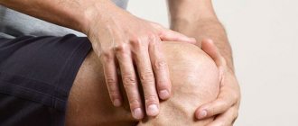
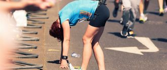
Knee injuries often occur when exercises are performed incorrectly.
The article will talk about traumatic injuries of the knee joint, their types, the clinical manifestations that accompany them, treatment and rehabilitation options, and also talk about the mechanisms of injury in the knee area to understand how they can be prevented.
Types of injuries
Classification of injuries and their causes:
| Name of knee injury | Possible causes, risk factors |
| Injury | Direct contact with a hard surface - impact, fall from the height of your own body |
| Medial ligament injury | Tucking the shin outward |
| Damage to the lateral ligament | Tucking the shin to the inside |
| Cruciate ligament injury | Flexion and extension of the knee under load with a large amplitude during sports activity (sprinting, bench press) Complex movement, for example, turning the hip when the lower leg is motionless, striking directly to the knee Commonly found in runners, hockey players, wrestlers, figure skaters |
| Injury to the patellar ligament | Bend the knee sharply if the thigh muscles are tense Kick, jumping The last stage of tendonitis of the patellar tendon (the disease occurs with constant increased loads on the joint) Chronic diseases that weaken the ligament (rheumatoid arthritis, diabetes, systemic lupus erythematosus) |
| Quadriceps tendon rupture | Sudden flexion of the shin if the thigh muscles are tight Hitting the foot on a hard surface |
| Meniscus injuries (tears, tears, flattening) | Strong physical activity Landing when jumping from height to feet Severe stage of knee arthrosis |
| Patella fracture | Falling with a blow to the front of the knee (sometimes even from one’s own height) |
| Fractures of the condyles of the femur or tibia (condyles are thickenings of the bone on which muscles are attached) | Falls from height Accidents or other traumatic situations |
Anatomical structure of the knee. Click on photo to enlarge
Pain in the back of the knee
Let's look at the most common causes of back knee pain (popliteal pain), less common causes, and major injuries that shouldn't be overlooked. The most common injury that causes posterior knee pain is hamstring (biceps, semitendinosus, and semimembranosus) tendonitis due to overuse, although other injuries and conditions, such as deep vein thrombosis, may also be potential causes of pain.
- Calf tendonitis
- Biceps tendinitis
- Baker's cyst
- Calf muscle rupture
- Damage to the popliteus muscle
- Posterior cruciate ligament rupture
Symptoms: general and depending on the injury
Different injuries present slightly differently, but the main symptoms are similar:
- Pain.
- Mobility disorders.
- Swelling.
- Swelling - sometimes.
If these symptoms occur, you should provide first aid to the victim (or yourself) and contact a traumatologist.
Signs of various injuries:
| Name of knee injury | Symptoms |
| Injury | Sharp pain immediately after the blow Gradually it becomes aching, low-intensity, but very intrusive Pain increases during physical activity and when the knee is touched Hematoma is also possible Sometimes, against the background of a bruise, synovitis develops - inflammation of the synovial bursa of the knee |
| Medial ligament injury | Severe pain during the injury itself In the future - swelling, pain when touching the injured knee, active movements involving the knee joint, especially if the lower leg is tilted outward |
| Damage to the lateral ligament | With sprains and tears - pain in the knee, especially in its outer part, limited movement, swelling, increased pain when the tibia deviates to the inside When a rupture occurs, too much mobility of the joint is added to the listed symptoms. |
| Cruciate ligament injury | High-intensity pain, clicking (if the anterior cruciate ligament is torn; if the posterior cruciate ligament is injured, it is usually absent) In the future: instability of the joint, hyperextension, pain and feeling of instability when walking, swelling, increase in the size of the joint With an old injury, the instability of the functioning of the joint is preserved, and frequent dislocations occur. |
| Damage to the patellar ligament | Pain, click In the future: pain and increased sensitivity in the knee, the joint is not able to fully straighten, it can “fall through” when walking, the kneecap moves upward as its fastening is disrupted |
| Quadriceps tendon rupture | Sharp pain, swelling just above the knee, which quickly increases. In the future: pain of moderate intensity just above the knee, limited knee movements, inability to walk normally |
| Meniscus injuries (tears, tears, flattening) | In the first time after injury: pain, swelling, limitation of movement After 2–3 weeks: periodic blockade of the joint (impossibility of movement), pain when walking down the stairs, staying in the lotus position, rotating the shin if the leg is bent at the knee Against the background of injury to the meniscus, synovitis often develops - inflammation of the synovial bursa, which is accompanied by the accumulation of fluid. |
| Patella fracture | Immediately after the fracture: severe pain, increasing swelling In the future: the patient cannot stand on his leg normally (intense pain occurs), cannot support his raised straight leg. |
| Fractures of the femoral or tibia condyles | Acute pain, severe swelling Deformation of the knee joint, inability to move it |
Pain in the front of the knee
Knee pain in the front may involve the kneecap or kneecap. The two most common causes of pain in the anterior kneecap are patellofemoral syndrome and patellar tendinitis. Sometimes it is difficult to say which of these two reasons is occurring, and sometimes they can occur simultaneously. The following are the main causes of anterior knee pain.
- Patellofemoral pain syndrome
- Patellar tendonitis (jumper's knee)
- Suprapatellar bursitis
- Osgood-Schlatter disease (osteochondropathy of the tibial tuberosity)
- Sinding-Larsen-Johanson disease (osteochondropathy of the patella)
- Chondromalacia patella
- Prepatellar bursitis
- Patellofemoral instability (patellar instability)
- Quadriceps tendinitis
- Infrapatellar bursitis
- Hoff's disease (inflammation of adipose tissue)
First aid
Almost any injury to the knee joint is accompanied by hemarthrosis - intra-articular hemorrhage. Proper care is designed to prevent hemarthrosis from progressing. Actions are also aimed at reducing pain.
Click on photo to enlarge
First aid instructions:
- Sit down and try not to move your knee.
- Apply a cold compress to the joint area: on the front and sides. This will help reduce pain and prevent an increase in intra-articular hematoma.
However, never apply cold to the back of your knee. This can lead to spasm of the popliteal vessels, which supply blood to the entire leg.
Possible consequences
Most complications develop as a result of the victim’s failure to seek medical help. The ligaments do not fuse correctly, which causes inadequate stabilization of the knee structures. They wear out faster - cartilage is destroyed, the bone surfaces of the tibia and femur change pathologically. Deforming osteoarthritis (gonarthrosis) develops, often leading to disability.
Diagnostics
When you go to the emergency room, your doctor will do the following:
- Ask the patient about symptoms.
- Feels the knee.
- Perform simple tests (e.g. leg elevation, flexion, extension, etc.).
- Prescribe an x-ray (if a fracture is suspected) or an ultrasound of the knee joint (for suspected injuries to ligaments, tendons, menisci).
If it is necessary to clarify the diagnosis, an MRI, CT scan, or arthroscopy may be needed (a procedure in which all manipulations in the joint - therapeutic and diagnostic - are done through small tissue punctures).
Technique for knee arthroscopy
general information
Knee ligament rupture is a common injury that occurs at home or during sports. These connective tissue strands are dense, strong, but not elastic enough. Therefore, when exposed to loads exceeding their tensile strength, partial rupture or complete separation from the bone base occurs, often with a bone fragment.
Treatment methods
Many injuries are complicated by hemarthrosis and synovitis (inflammation of the synovial membrane surrounding the joint), so for treatment, a puncture (tissue puncture to penetrate the joint) is performed to remove accumulated fluid from the joint.
Other treatments depend on the type of injury.
Knee joint injuries and their treatment:
| Type of damage | Conservative treatment methods | Surgical treatment methods |
| Injury | First day – cold compresses Next until recovery - UHF, rest | Not required |
| Sprains | Wearing a special bandage that relieves excessive stress and speeds up recovery | Not required |
| Ligament tears | Gypsum bandage After its removal - massage, therapeutic exercises | Not required |
| Ligament tears | Not effective | Sewing the ligament or its plastic |
| Tendon rupture | Not effective | Stapling operation |
| Meniscus injury | Effective for minor injuries A plaster cast is applied and rest is recommended. After removal, therapeutic exercises, physiotherapeutic procedures and drug treatment are prescribed:
| If the injury is severe or is accompanied by recurrent synovitis, then surgery is prescribed Depending on the severity of the injury, 4 types of surgical interventions are possible: suturing of the meniscus, resection (cutting), complete removal, transplantation (removal and replacement with an artificial one) |
| Patella fracture | Gypsum bandage | If the fracture is displaced, then surgery is performed During this process, the fragments are connected to each other using a special wire. |
| Fracture of the condyles of the leg or femur | Skeletal traction | If displacement occurs, then insertion of a plate, screws or special bolts into the bone is necessary. |
On the left is a knee brace, on the right is a plaster cast. Click on photo to enlarge
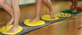
Rehabilitation
Rehabilitation measures are necessary after a long stay of the leg in a cast, as well as after operations.
These include:
- therapeutic exercises;
- massage treatments;
- physiotherapy;
- gentle regime of physical activity;
- sometimes - medications (chondroprotectors that protect cartilage: Dona, Structum, Elbona; as well as NSAIDs that prevent inflammation: Ibuprofen, Ketoprofen, Piroxicam).
After severe injuries, an athlete may be advised to take a long break or retire from professional sports.
Click on photo to enlarge
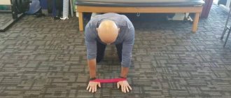
Examples of exercise therapy exercises for rehabilitation of the knee joint. Click on photo to enlarge
Prognosis for knee injury
Different injuries heal at different rates:
- Full recovery after a bruise occurs within 2 weeks to a month.
- A sprain goes away in 2 weeks.
- If the ligaments are torn, a cast is applied for 3–4 weeks, then rehabilitation measures are carried out for several more weeks.
- Meniscus injuries can be treated over several months. You need to protect the joint from excessive stress for another 6–12 months.
- A fracture of the patella heals in 1.5–2 months, during which the patient wears a cast. Then rehabilitation measures are prescribed for another 2–4 weeks. The entire recovery process takes 2–3 months.
- Full recovery from condyle fractures takes up to 1 year.
The main thing is to contact a traumatologist in time, since an old injury can cause pain for many years after healing.
Knee contusion
Knee bruise
- this is damage to surrounding tissues (skin, subcutaneous fatty tissue), which is not accompanied by a violation of the anatomical structure. Against the background of this injury, inflammation of the elements that are located inside the joint occurs, as well as hemorrhages. Symptoms are often not as pronounced with other injuries. Because of this, the diagnosis is made when other injuries have been ruled out. Signs that the victim has a knee joint injury may include: pain, moderate swelling of the soft tissues, and slight bruising. At the same time, support is maintained, but the person may begin to limp and be slightly limited in some movements.
To determine pain, palpation is performed in the area of the bruise. Feeling the bones and ligaments should not cause attacks of pain, as well as pathological mobility. There may be an accumulation of fluid, at first it is blood, and after two or three weeks it is exudate. Injury to the knee structures is excluded using x-rays. Research methods such as MRI, computed tomography, ultrasound and arthroscopy are also used. Care for a knee injury is provided at the emergency room. If there is synovitis or blood inside the joint, a puncture is performed. If the patient has a slight bruise, then the main actions are aimed at maintaining peace. But for severe cases, the traumatologist applies a plaster cast for 2 - 3 weeks. For the first two days, it is recommended to apply something cold to the site of the bruise, and from the third day, UHF therapy is prescribed. The patient must visit the doctor at the appointed time and follow his instructions. If indicated, repeat arthrocentesis is performed. If the knee joint is bruised, the disability lasts 2–4 weeks.