The glenohumeral joint is a ball-and-socket joint. This is one of the four joints that make up the shoulder complex. It is formed by the head of the humerus and the glenoid cavity of the scapula. The glenohumeral joint is considered the most mobile, least stable and most susceptible to dislocation.
Movements in the glenohumeral joint
Planes in anatomy
- Abduction (abduction) - raising the shoulder in the frontal plane.
- Flexion (flexion) - raising the shoulder anteriorly in the sagittal plane.
- Extension (extension) - raising the shoulder posteriorly in the sagittal plane.
- Internal rotation (internal rotation) - rotation of the shoulder inward (in the medial direction).
- External rotation (external rotation) - rotation of the shoulder outward (in the lateral direction).
- Abduction in the plane of the scapula is the elevation of the shoulder in the plane of the scapula, which is between the frontal and sagittal planes.
- Horizontal adduction is the inward movement of the shoulder in the horizontal plane (usually accompanied by some degree of shoulder flexion).
Joint capsule and ligaments
The joint capsule and ligaments of the glenohumeral joint provide passive support for the head of the humerus in contact with the glenoid cavity of the scapula.
Capsule of the glenohumeral joint
Capsule of the glenohumeral joint
- Laterally, the capsule of the glenohumeral joint is attached to the anatomical neck of the humerus.
- Medially, the capsule is attached to the glenoid cavity and the articular labrum.
- When the arm is in a resting position, the lower and anterior portions of the capsule are relaxed while the upper portion is taut.
- The anterior portion of the capsule is strengthened by the superior, middle, and inferior glenohumeral (glenoid-shoulder) ligaments, which form a Z-shaped pattern on the capsule (some sources call this the articular-labial periarticular fibrous complex).
- The rotator cuff muscles strengthen the joint capsule superiorly, posteriorly, and anteriorly.
- Without ligaments and surrounding musculature, the joint capsule provides little support to the glenohumeral joint.
- According to some sources, the overall strength of the capsule has an inverse relationship with the patient’s age: the older the person, the weaker the joint capsule.
Shoulder, shoulder girdle and shoulder joint[edit | edit code]
The shoulder girdle consists of the collarbone and scapula. The proximal end of the clavicle forms with the sternum the sternoclavicular joint, the only joint connecting the shoulder girdle with the axial skeleton. This joint is strengthened by the anterior and posterior sternoclavicular, costoclavicular and interclavicular ligaments. The distal end of the clavicle and the acromion form the acromioclavicular joint, which is strengthened by the coracoclavicular and acromioclavicular ligaments.
The glenoid cavity of the scapula and the head of the humerus form the shoulder joint. This is a very mobile and therefore rather fragile spherical joint, reinforced by the articular labrum, articular capsule and articular-brachial ligaments.
Movements of the arm in the shoulder joint (Fig. 5.1) occur under the action of many muscles. Flexion is carried out by the clavicular part of the pectoralis major muscle and the anterior part of the deltoid muscle. Extension occurs due to contraction of the latissimus dorsi muscle, teres major muscle and the sternocostal part of the pectoralis major muscle. Abduction is provided by the deltoid muscle and the rotator cuff muscles (subscapularis, supraspinatus, infraspinatus and teres minor). Adduction occurs due to the contraction of the pectoralis major muscle (sternocostal part), the latissimus dorsi muscle and the teres major muscle. The subscapularis and pectoralis major muscles internally rotate the humerus, while the supraspinatus, infraspinatus and teres minor muscles externally rotate the humerus. Horizontal adduction is accomplished by simultaneous contraction of the coracobrachialis, pectoralis major, and anterior deltoid muscles, and horizontal abduction is accomplished by contraction of the infraspinatus, teres minor, and posterior deltoid muscles.
Figure 5.1.
Range of motion in the shoulder joint.
A.
Flexion and extension.
B.
Abduction and adduction.
B.
Rotation outwards and inwards.
D.
Horizontal abduction and adduction.
Figure 5.1
(end). Range of motion of the scapula.
D.
Raising and lowering.
E.
Outward and inward rotation.
G.
Abduction and adduction.
The rotator cuff is the muscles immediately adjacent to the capsule of the shoulder joint (subscapularis, supraspinatus, infraspinatus and teres minor).
Simultaneously with the movements in the shoulder joint, movements of the scapula occur, namely its abduction, adduction, rotation outward or inward, as well as elevation and descent. Abduction of the scapula is carried out by the pectoralis minor and serratus anterior muscles, adduction by the rhomboid muscles, rotation of the lower angle outward by the serratus anterior and trapezius muscles, rotation of the lower angle inward by the pectoralis minor and rhomboid muscles, elevation by the levator scapulae muscle, and descent by the pectoralis minor muscle. .
Bursae of the shoulder joint
Bursae of the shoulder joint
The shoulder complex has many bursae, the largest being the subacromial bursa. It also includes the subdeltoid bursa as they are often continuous. The subdeltoid bursa allows the rotator cuff to slide easily under the deltoid muscle.
In order not to miss anything interesting, subscribe to our Telegram channel.
Adduction of the shoulder at the shoulder joint
Posterior muscle group of the shoulder girdle.
Every load carried, pulled or pushed by the hands transfers its weight to the axial skeleton and then to the ground through the shoulder skeleton and musculature.
You can't lift a weight off the ground without using your shoulder muscles. You can't push anything without stressing your shoulder. Even something as simple as scratching your forehead requires relative tension in your shoulder muscles. Shoulder muscles can be divided
by layers:
tangible superficial muscles and intangible deep muscles.
by location:
front, rear and side.
The posterior muscles include the trapezius, movator scapulae, rhomboids, latissimus dorsi, triceps, biceps, serratus anterior and deltoids.
Note that some muscles have anterior, lateral, and posterior aspects.
Figure 1: Posterior muscles of the shoulder: trapezius, movator scapula, rhomboid, and posterior deltoid.
Trapezoid
- is a large and recognizable superficial muscle of the upper back. Its geometric shape is roughly trapezoidal (hence the name).
Vertically stretches from the occipital protuberance to the level of the lower thoracic vertebrae.
Laterally from the vertebral processes to the scapula.
The important observation here is that the spinal column remains stable during most movements. This means that the scapulae generally move relative to their attachment to the axial skeleton: they move towards and away from the axial skeleton, rather than vice versa.
The upper trapezius lifts the shoulder blades and therefore the shoulders, allowing the shoulders to rise upward.
The middle trapezius pulls the shoulder blades toward the center, causing retraction.
The lower trapezius pulls the shoulder blades down, lowering the shoulders.
Diamond-shaped
- has two parts (large and small), shaped like a rhombus.
The small one is smaller and is located above the large one. Although this muscle is made up of two separate parts, it generally functions as a single unit.
Located under the trapezius muscle, the rhomboid minor is attached to the spinous processes of the seventh cervical vertebra and the first and second thoracic vertebrae. The rhomboid major is attached to the spinous processes of the second, third, fourth and fifth thoracic vertebrae. Both the major and minor are attached to the medial border of the scapula at approximately the level of the scapular process, with the attachment of the major extending to the inferior angle of the scapula.
Contraction of the rhomboid muscle usually causes retraction of the shoulder blades (pulling them closer to the central axis of the body and squeezing the shoulder blades together). The diamond major helps hold the shoulder blades (and therefore the upper limb) to the ribcage. It also rotates the scapula downward in relation to the shoulder joint and works synergistically with the scapular adductor muscles to elevate the medial border of the scapula.
Muscles that move the scapula
- These are muscles located deep and below the trapezius muscle. They are attached to the transverse processes of the Atlas and the third and fourth cervical vertebrae. Distally, it attaches to the superior medial border of the scapula.
When the axial skeleton and skull remain stable, it is the load-bearing muscle that elevates the medial scapula.
In short, she will get tired while hiking with a heavy load in her backpack.
Figure 2: Latissimus dorsi and triceps deltoid muscles.
Latissimus
("latissimus" - Latin for "widest") - many people think of it as a very broad, very large superficial back muscle that gives the upper body a V-shape, in fact it makes a solid contribution to the work of the shoulder and performs five functions of shoulder motion: extension, adduction, horizontal abduction, extended flexion, and internal rotation. The muscle also promotes extension and lateral flexion of the lumbar vertebrae.
Muscles of the glenohumeral joint
Muscles of the shoulder complex
Flexors
- anterior portion of the deltoid muscle.
Extensors
- triceps brachii;
- teres major;
- posterior portion of the deltoid muscle;
- latissimus dorsi muscle.
Rotator cuff muscles
- supraspinatus muscle;
- infraspinatus muscle;
- teres minor;
- subscapularis muscle.
Internal rotators
- subscapularis muscle;
- teres major;
- latissimus dorsi muscle;
- pectoralis major muscle.
External rotators
- teres minor;
- infraspinatus muscle.
Abductors
- deltoid;
- supraspinatus muscle.
Adductors
- pectoralis major muscle.
Exercises and technique
All exercises can be performed at home, in the gym (you can attach the expander to the stand of the exercise machine) or on an outdoor sports ground (you can attach it to the support of the horizontal bar). When doing exercises at home, provide in advance places on the walls where one end of the expander or the middle part of the spring can be firmly secured.
We will talk about analogues of presses and flyes.
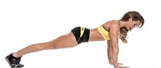
Press with expander
The exercise is best performed while standing, because for the lying version you will need a bench with a variable backrest angle. You won't be able to perform the bench press on the floor because your elbows go below the floor during this exercise. You will not be able to make full range of movements. That's why we do everything standing.
- Stand straight with your feet shoulder-width apart. Straighten your shoulders, bend your lower back. To control, approach the wall with your back and press against it so that your heels, buttocks, shoulder blades and the back of your head touch the wall. This is correct posture with all natural deflections. Remember this pose, take 2-3 steps forward.
- If there is nowhere to fasten the spring, place it on your back at the level of the lower part of your shoulder blades and straighten your arms forward, holding the handles. If you raise the spring higher, it can jump onto your neck. Palms turned down. Starting position – arms in front of you, expander stretched. If you can secure the middle of the spring behind you at the level of your shoulder blades, it will be more convenient.
- We begin to bend our arms, orienting our elbows strictly to the side. Elbows should move parallel to the floor. The spring should pull your hands in that direction.
- Move your elbows back as far as possible, stretching the pectoral muscle. Now hold this position for 1-2 seconds.
- Straighten your arms to the starting position.
Do 10-15 repetitions in 3-4 sets. It’s difficult to work on mass here, but on relief it’s quite possible. If you have a lot of strength left, you can do 30 repetitions. The endurance of the muscles of the chest, arms and shoulders will increase.
It is important to watch your elbows in this exercise. If performed correctly, the pecs and triceps will work. Since the exercise is done standing, the shoulders will receive a good load.
If you have an inclined bench at home, you can repeat the same thing while lying down, throwing the expander behind the bench. It will be more convenient and familiar. Change the backrest to an angle of 30, 45 and 60 degrees. At the same time, straighten your arms straight up. Then you will pump up the entire muscle mass.