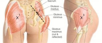
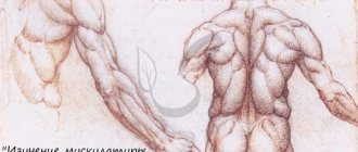
The pectoral muscles (pectoral from the English pectoralis) are one of the main objects of training in strength sports, both for men and women. At first glance, they have a simple structure and function; up to 90% of the entire group is represented by the pectoralis major muscle. However, the thoracic region includes several components. Understanding their characteristics can make chest training as productive as possible.
The structure of the pectoral muscles
The entire area is represented by the following chest muscles:
- Large;
- Small;
- Serrated;
- Subclavian.
The latter is the smallest in volume in the group, therefore almost no attention is paid to it in sports.
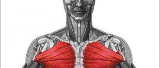
If we take into account the anatomy of the pectoral muscles, the large one occupies almost the entire volume of the group. The structure of this pectoral muscle has a complex structure. The muscle fibers are directed in different directions, therefore, to work out the entire surface, a load is required from different angles. The pectoralis major starts from:
- medial part of the clavicle;
- anterior wall of the rectus abdominis muscle;
- anterior surface of the sternum and costal cartilages.
Attaches to the crest of the greater tubercle of the humerus and the edge of the deltoid. Structurally, it is the largest muscle of the chest, which covers the area in the form of a shield or solid plate.
The pectoralis minor muscle is the second largest in the group. It lies under the large one and starts from 2 to 5 costal teeth. Attached to the scapula (coracoid process).
The serratus pectoralis muscle is anatomically located away from the main group. Like the small one, it starts from the ribs and is attached to the shoulder blade.
The subclavian starts from the 1st rib (cartilaginous part) and is attached to the lower part of the clavicle.
In addition to the main group, the rib muscles are distinguished. They belong to infants and are divided into:
- superficial chest muscles;
- as well as the deep muscles of the chest.
They are responsible for inhalation and exhalation; they usually do not need additional training.
Anatomy of the female breast
The chest is a paired organ located at the level of 3–6 ribs. The bust is attached to the anterior pectoral muscle, but there are no muscles in the gland itself. The breast consists of glandular, fatty and fibrous tissue. Its structure is tubular-alveolar.
Reference! Alveolus - from the Latin word alveolus “cell, depression, vesicle.” In the case of glandular tissue, the alveolus is the cell-shaped end of the gland.
The glandular body is formed from separate parts - lobes. There can be from 15 to 25, depending on the individual characteristics of the woman. The lobe has the shape of a cone, with its apex facing the nipple. All cone-shaped parts are located around the nipple along its entire radius.
The nipple is surrounded by the areola - a pigmented isola. Its most convex part has pores through which milk flows.
The body of the gland is enclosed in a capsule of fatty and connective (fibrous) tissue. The same type of tissue insulates the lobes from each other.
The layer of subcutaneous fatty tissue that immediately follows the skin and surrounds the entire gland is called premammary. Premammary fat is absent in the areola area. Fat cells that separate the lobes are called intramammary.
The mammary gland is attached to the chest wall by connective tissue (fascia). Following the fascia is a layer of fat. It forms a kind of “pillow” on which the bust rests.
Glandular tissue
As mentioned, the mammary gland is divided into lobes. Each lobe in structure resembles a bunch of grapes and consists of “grapes” - alveolar glands with a diameter of 0.05–0.07 mm. Alveoli are secretory units. That is, during lactation, each woman produces milk, which is excreted through tubes - the milk ducts. Ducts “branches” connect the alveoli to each other. Along the way, they gather into wider tubes and rush towards the nipple, forming a spindle-shaped expansion in front of it - the milk sinus. The diameter of the ducts is 1.7–2.3 mm. The terminal part of the milk sinus is narrowed, it penetrates the thickness of the nipple and opens outward with the milk opening. There are 15–18 such holes.
Nipple
Its color and size vary from person to person, but usually in nulliparous girls it is pink or dark red. After childbirth, the nipple often darkens, acquiring a brownish color.
The nipple and areola have many nerve endings. This allows the mammary gland to sensitively respond to the touch of the baby's mouth during feeding, responding to it by increasing milk production. This increased sensitivity of the nipple makes it an erogenous zone.
Basic functions of the chest muscles
In most cases, the functions of the pectoral muscles are duplicated, the entire array works as a single whole. Therefore, in sports, the entire group is linked by the function of the pectoralis major muscle, without isolating the rest separately.
Pectoralis major muscle:
- lowers his arms to his body from the top position;
- pronation (turning inward) of the hand;
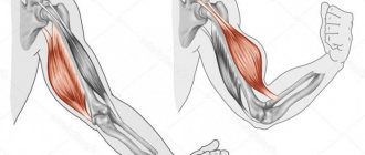
- flexion of the arm (responsible for the clavicular part);
- assistance with inhalation and pulling the body (for example, while climbing a rope).
Pectoralis minor muscle:
- provides movement of the scapula;
- pulls the scapula up and down;
Also, the functions of the pectoralis minor muscle include assisting in breathing with fixed arms.
Serratus muscle:
Rotating and pushing the shoulder blade forward.
Subclavian muscle:
Provides downward movement of the collarbone.
In men and women, the chest muscles anatomically have almost no differences, with the exception of some features. The only visual difference is the mammary glands. And also in the anatomy of the pectoral muscles of men there is a predominance of fast muscle fibers. In women, the ratio is shifted towards slow fibers.
Exercises to develop chest muscles
To have good posture, you need to maintain muscle tone throughout the body. The pectoral muscles are no exception. It is important to remember that at the present stage there is a large amount of conflicting information about training the chest muscles. A modern person is required to clearly understand that the most important thing in training the pectoral muscles is the correct load on them.
Below are tips for training your chest muscles.
• It is not recommended to combine training of the chest and triceps muscles on the same day, because this leads to disruption of muscle adaptation to increased load.
• It is not recommended to carry out training exercises to strengthen the chest muscles more than twice a week.
• The sum of approaches to performing exercises for the chest muscles should not exceed 6.
• The active phase of chest muscle exercises should not be prolonged. Slow transitions from one set of exercises to another are necessary.
• The best sets of exercises for the chest muscles are those that involve push-ups.
• From an anatomical point of view, sets of exercises for the chest muscles are horizontal push-ups.
Let's name the most popular and effective sets of exercises for the pectoral muscles.
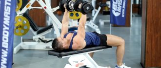
• Incline bench press
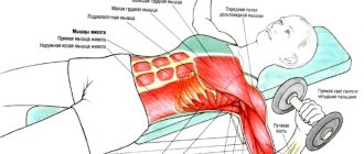
Lifting the barbell effectively affects all pectoral muscles. This exercise requires strict adherence to the rules for its implementation. It is imperative to perform the movements in full. The back of the bench should be at an angle of fifteen to thirty-five degrees. You need to sit so that the soles of your feet are wider than your shoulders. The head, shoulder girdle and hips should touch the bench. The grip on the bar must be correct. You need to inhale and lower the barbell to your chest. You need to exhale and lift the barbell up.
• Incline dumbbell press
Raising dumbbells on an incline bench has a wonderful effect on the relief of the chest muscles. The most important thing in this complex is a constant range of motion. It must be remembered that in this case some preparation is necessary: lower stretching. It is important to maintain an inclination of twenty to thirty degrees.
• Dumbbell flyes in a horizontal position
This exercise is less productive than those mentioned above, because the movements in it are somewhat closed. Its advantage is that when it is performed, high-quality pumping of the pectoral muscles occurs. The advantage of the exercise is that it contributes to the appearance of a beautiful relief of the pectoral muscles.
• Chest dips
This set of exercises can be called a classic for the chest muscles. It is important to remember that the most valuable thing in the named complex is the technique of its implementation. The technique includes maximum load on the pectoral muscles. It has a qualitative effect on the lower and middle sections of the pectoral muscles.
• Exercises for chest muscles with a ball
There are two such complexes.
— The essence of the first complex is that the ball needs to be thrown into the floor as if diagonally in relation to your own body. You must perform up to twenty throws each time.
— The second complex is push-ups with a ball. When doing push-ups, one of the hands should be on the ball. Each limb should perform this procedure up to ten times.
The best exercises for chest
- Bench press - works almost the entire shoulder girdle and arms.
- Bench press at an angle - focus on the upper chest.
- Dumbbell press (on a straight and incline bench) - duplicates the barbell press, but it loads the pecs to a greater extent.
- Lying dumbbell flyes.
- Pullover – loads the entire pectoral mass, especially the serratus muscle.
- Dips – focus on working the lower part of the pecs.
- Bringing the arms together in a crossover - working out the lower and inner parts.
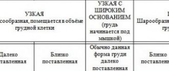
Size and shape
Women's breasts are generally larger in size than the mammary gland. The size of the bust largely depends on the content of fat cells in it. It’s not for nothing that if a girl loses weight, her breasts “lose weight” along with her - after all, fat leaves the body evenly.
As a rule, the breast size of thin petite women is small, while larger or fuller ladies have a solid bust. However, there are exceptions - sometimes it is the glandular tissue that develops intensively. In this case, a thin woman may have large or even very large breasts - this is called macromastia. Accordingly, underdevelopment of the glands is micromastia. It is expressed by the practical absence of a bust, regardless of the build of its owner.
Breast asymmetry occurs quite often - this is a normal variant. The difference between the right and left breasts can be up to two sizes.
The tendency to ptosis (sagging) depends on the elasticity of the connective tissue of the chest capsule, the shape and size of the bust. The heavier it is, the more it will stretch even the elastic capsule and, accordingly, sag. In most cases, appetizingly protruding breasts of size 5+ are the result of the use of implants. The tendency to ptosis depends not only on the size, but also on the shape of the bust.
Shape and size do not affect the health of the breast and its ability to lactation - this is a purely aesthetic issue.
Fact! The “chest exercise” recommended in women’s magazines is aimed at developing the anterior pectoral muscles on which it is attached. But these muscles have little effect on the fit of the bust and its shape, since it itself does not have muscle tissue. Therefore, if a woman is not satisfied with the appearance of her breasts, this can only be eliminated surgically.
Breast surgery can be performed no earlier than 18 years of age. And preferably after the first lactation, since surgery can affect the ability of the gland to produce milk.
Muscle strain
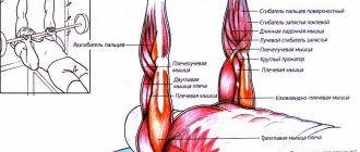
If a person is regularly exposed to physical activity on the pectoralis minor muscle, the process will lead to impaired functioning of the shoulder system. As a result, the risk of injury to this area as a result of lifting heavy objects or sudden movements increases significantly. The most common problem among patients is subacromial impingement syndrome. This is a condition in which there is not enough space for the muscle group between the humerus and the acromion. The disease leads to pinching of the tendon of the periosteum muscle, as well as the subacromial bursa. The syndrome is especially evident when lifting the limb above shoulder level and during rotational movement. Associated symptoms include the following:
- dull pain in the shoulder area;
- increased pain when raising your arms;
- crunching or clicking in the shoulder;
- limited mobility of the affected shoulder;
- weakness in the hand;
- problems with dreams due to pinched blood vessels and nerve fibers.
A negative process resulting from overstrain of the pectoralis minor muscle leads to pinching of nerve fibers, tendon tissue, arterial and venous vessels.
Patients are often diagnosed with hypertonicity of the pectoralis minor muscle. At the same time, the patient feels painful feelings. This is a condition in which when a muscle is loaded, it puts up a lot of resistance. This pathological condition leads to difficult movement and various symptoms. In order to restore physical activity, it is necessary to consult a doctor in a timely manner, undergo diagnostics and begin treatment. As a rule, patients are recommended therapeutic massages, physiotherapy, and gymnastic exercises. The sooner treatment is started, the easier it is to restore the body and prevent curvature of posture.
Hormones affecting the mammary gland
The mammary gland is the target organ for 18 currently known hormones. It responds to all changes in the complex hypothalamic-pituitary-ovarian system. The most significant and direct influence on it is exerted by female sex hormones - estrogen, progesterone and prolactin.
Estrogens
Sex hormones that begin to be produced by the ovaries with the onset of puberty. Under its influence, the formation of the mammary gland occurs - the growth of ducts, the appearance of alveoli at their ends. The formation of connective and adipose tissue increases. The breasts take on a mature shape. However, the mammary gland is fully formed only during pregnancy.
Under the influence of this hormone, the breasts become fuller and thicker before menstruation.
Progesterone
The hormone is responsible for the development of glandular tissue and its receptors. With the onset of pregnancy, the amount of progesterone increases sharply, and under its influence the alveoli are finally formed. Modern science does not yet have a complete picture of the interaction between progesterone and the mammary gland.
Prolactin
It has a great influence on the preparation of the mammary gland for lactation during pregnancy and regulates the lactation process itself. In the third trimester of pregnancy and during breastfeeding, the amount of prolactin increases significantly. With the combined influence of prolactin and progesterone, the growth of mammary gland cells becomes 3-17 times more intense.
Prolactin not only stimulates milk production, but also affects its nutritional value.
Breasts during menopause
With the onset of menopause, ovarian function dies out, female sex hormones cease to support the development of glandular tissue. It gradually begins to be replaced by fatty and fibrous tissue. This process is called breast involution.
Functional meaning
The superficial muscles of the sternum in the human body are represented by several types of groups, among which the following types stand out:
- pectoralis major muscle;
- pectoralis minor;
- serratus anterior;
- subclavian
One of the largest and strongest groups is the pectoralis major muscle. Its location covers a fairly large part of the chest from the front. The shape of the group resembles a fan, the beginning of which is located at the armpit, and the wide part is located in the center of the sternum. It is flat and steamy. The main functional tasks are the following functions:
- Lowering the arms from the position of raised limbs;
- Bringing the arms to the body from the position of raised limbs;
- Performs limb rotation;
- Participates in the respiratory process to expand the chest;
- Allows you to raise the ribs with well-fixed limbs.