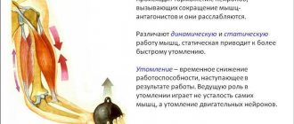
Separation of leg muscles
The lower body has more than 30 muscles. But we are not interested in everyone. Since we are not trying to pass the medical examination. Our goal is to develop the core muscles that will help us achieve outstanding results in the gym. And also, give shape to the lower part of our body. Anatomically, we can divide the leg into 4 parts:
- Pelvis (buttocks)
. These muscles are not exactly part of our leg. But since they are attached to the femur, we take them into account too. And it’s very difficult not to engage your glutes when performing most leg exercises. This muscle group is located on the back side of the body. In the area between the lower back and thigh. Of greatest interest to us are: the gluteus maximus, the gluteus maximus and the gluteus minimus. - Hip. This is the part that almost all athletes want to develop. It is what gives our legs their shape. It starts from the pelvic bone (to which some muscles are attached) and ends at the knee. Its main functions include flexion and extension of the lower leg and thigh. As well as bringing the legs to the body and their rotation (rotation). Conventionally, this group is divided into parts. Anterior, medial (internal) and posterior.
- Shin. These are not large, but very durable muscles that occupy the lower part of the leg. From knee to foot. We are interested in the back group. This includes the triceps and plantaris muscles. All other muscles are responsible for flexion and extension of the fingers. Therefore, we will not consider them.
- Foot.
This is our support. Which is responsible for balance. The muscles located in the foot mainly start from the lower leg. And they are responsible for the movement of fingers. Their flexion and extension. As well as sideways movement relative to each other. And for the turns of the foot. When performing any exercise that requires balancing the body, they are actively involved. Therefore, we will not do separate exercises for these muscles. Still, our task is to devote most of our time to the larger groups.
We can also divide the leg muscles according to the degree of their location:
- Superficial . These are the muscles that are located in plain sight directly under the skin. We can easily touch them with our hands. These muscles give that visual appearance.
- Deep. Located under the surface. And even though they are practically invisible. But they also play a huge role in the formation of legs.
But don't forget one more point. When training our legs, we not only strive to give them one shape or another. We also face the task of making these muscles stronger. This will increase the stability of our body and we will be able to lift heavier weights. And for people involved in strength sports, load progression is one of the key factors.
Features of the structure of the leg muscles
Unlike other muscle groups, the legs have the greatest number of functions. This is the largest collection of muscles that are closely intertwined with each other. Everything is aimed at stabilizing and providing maximum functionality to the three pairs of joints that interact with the muscles of the human legs:
- hip;
- knee;
- ankle
A special role in ensuring human life is played by the muscles of the lower leg: gastrocnemius and soleus. These are powerful pumps that are directly involved in blood circulation. Because of this, calves are often called the “second heart.” Therefore, you need to train your legs not only to increase muscle size or endurance, but also to improve your overall health . Moreover, given the number of muscle fibers and the overall anatomy of the leg muscles, the lower body is designed for heavy workload. This allows you not only to work with heavy weights, receiving a powerful anabolic response, but also to significantly increase your overall endurance (which is more than 50% dependent on the legs).
In sports, the lower body is divided into 4 areas, taking into account their main functions:
- buttocks;
- muscles of the front of the thigh;
- muscles of the back of the thigh;
- calf muscles.
Leg muscles diagram-drawing
For productive training, there is no need to know the names of all leg muscles, taking into account each individual muscle. This is more related to medical topics. However, in order to avoid injury and increase the effectiveness of each exercise, it is necessary to at least generally understand the structure of the muscles of the human legs and the task of each individual area.
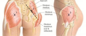
Buttocks
From the point of view of the anatomy of the human leg muscles, the gluteal group is considered one of the largest. Moreover, it includes the most massive muscle in the body – the gluteus maximus. The entire area is formed by three gluteal muscles:
- big;
- average;
- small
If you look at the general diagram of the leg muscles, the gluteal structure is designed in such a way that the muscles cover each other in layers, providing mobility and protection to the hip joint. The functions of the gluteal muscles include:
- Straightening the body;
- Taking the leg back;
- Taking your leg to the side.
Gluteal muscles
Front thigh
The entire anterior part is largely represented by a single large muscle, which consists of four heads. From the point of view of the anatomy of the human legs, the muscles and ligaments of the quadriceps are considered one of the strongest. Thanks to this, even beginner athletes can use impressive weights in squats, far exceeding the weight of weights in upper body exercises.
The quadriceps has 4 bundles:
- lateral (outer);
- medial (internal);
- average (intermediate);
- rectus femoris muscle.
The main function of the muscle is to extend the leg at the knee, although the quadriceps is also directly involved in bending forward and flexing the hip.
In addition to the quadriceps, the sartorius muscle also belongs to the front of the thigh. It is considered the longest muscle in the body and runs over the quadriceps.
Leg muscles: anterior group
Posterior thigh
Another large muscle area, which is represented by three muscles. If you look at the picture of the anatomy of the leg muscles, the bulk of the back of the thigh is occupied by the biceps muscle (aka “biceps femoris”), which includes two heads:
- long;
- short.
The main function of the biceps muscle is torso extension (works in tandem with the gluteal muscle).
Also included in the back of the thigh are:
- Semimembranosus muscle - responsible for hip extension, flexion and rotation of the lower leg.
- Semitendinosus muscle - performs the same functions as the semimembranosus.
If we evaluate the general purpose of the flexor and extensor muscles of the leg, the main task of the entire posterior surface is flexion of the leg at the knee joint (as well as rotation) . The muscles are also involved in hip abduction and external rotation.
Posterior leg muscles
Adductor muscles
This muscle group is located primarily on the inner thigh. Its main and only function is adduction and supination of the hip. The group includes 5 muscles that form a tightly woven bundle:
- Adductor magnus;
- Long adductor;
- Short adductor;
- Comb;
- Thin.
Leg adductors
This muscle group receives the least attention in fitness, although it plays an important role. If the hip adductors are underdeveloped, they can become a limiting factor in other exercises.
Calf muscles
All muscles of the lower leg are represented by two groups: anterior and posterior. The posterior includes three main and largest muscles , which are closely intertwined into a single bundle:
- Gastrocnemius (aka biceps) - includes the lateral and medial heads. This is the largest muscle below the knee.
- Soleus – located under the gastrocnemius muscle. Athletes who do not know what muscles are in their legs often ignore working the soleus muscle, although it is responsible for the volume of the calves.
- The plantaris is a muscle with long tendons that performs the same functions as the soleus and gastrocnemius. It is considered insignificant and may be completely absent (approximately every tenth person does not have it).
The front part of the lower leg is represented by the tibialis anterior muscle, the main task of which is supination and extension of the foot.
If we evaluate the lower part of the leg muscles taking into account the anatomical description, the main functions of the group include:
- flexion of the ankle and foot;
- supination and extension of the foot;
- rotation of the lower leg (inward).
Leg muscles below the knee
Anatomy of the skeletal bones of the lower limb (leg, pelvis)
Before we start looking at the leg muscles, we'll talk a little about the bones they are attached to. We won’t go too deep, since this is the topic of a completely different article. Below in the picture we can see the skeleton of our lower part.
As you can see, it all starts from the pelvis. Which consists of two iliac bones. The muscles of the pelvis and thigh are attached to their edges, namely the iliac crest. There is also a sacrum, which is attached to the spine. It allows us to keep our body upright. And already at its end, there is a coccyx. Below are two pubic bones. The edges of which serve as the attachment point for the adductor muscles and the back of the thigh. Now let's move on to the leg bones. The largest of them are the femoral ones. At their ends there are two so-called heads, which are attached to the ilium. Next to these heads, there are two protrusions, these are the greater and lesser trochanters. At the lower ends of the femur, there are two epicondyles. Medial (internal) and lateral (external). Also, there are two bones in our lower leg. Tibial and fibular. At the top of the tibia, located are two condyles. Medial and lateral. They serve as places for muscles to attach to them. All the bones of the leg connect to form the knee. There is a small bone there called the kneecap. Better known to everyone as the kneecap. And of course, don’t forget about the bones of the foot. Where are the phalanges of all our fingers located? And the large calcaneus.
This information will be enough to give you an idea. And it will be easier for you to perceive further information.
Muscles of the pelvic region
As I said earlier, this includes the gluteal muscles. For the sake of the development of which, a large number of girls in the hall shed a lot of sweat. But this does not mean that men avoid training them. It’s just that their priority is not the round shape, but the strength of these muscles. For example, in powerlifting and weightlifting, the development of the gluteal muscles is given great importance.
Gluteus maximus
It is a very large (hence its name) and flat muscle. The shape is vaguely reminiscent of a rhombus. Its development among all gluteal muscles is a priority. It is located on the surface and covers all the other muscles of this group. At the top it is attached to the posterior surface of the ilium. And also to the lateral edge of the sacrum and coccyx. Directing obliquely downward with its upper bundles, it is woven into the fascia lata (the protective sheath of the muscles). And the lower ones are attached to the gluteal tuberosity of the femur.
Functions:
Extends the hip, abducts it to the side and is responsible for its outward rotation. With fixed legs, rotates the pelvis.
Gluteus medius
This muscle is located immediately under the gluteus maximus. The shape resembles a triangle. Attaches to the outer surface of the ilium wing. Heading down, it turns into a powerful tendon. And is attached to the outer surface of the greater trochanter of the femur.
Functions:
Extends the hip. Participates in hip abduction and is the most powerful muscle that performs this movement. The position of the pelvis stabilizes when we stand on one leg. Also involved in lateral and medial rotation (outward and inward rotation).
Gluteus minimus
This is the smallest and deepest muscle of the three gluteal muscles. The top is covered by the middle one and completely resembles its shape. Just a smaller size. And it is attached in the same places. Above to the outer surface of the iliac wing. And below to the outer surface of the greater trochanter of the femur.
Functions:
Together with the gluteus medius, it moves the leg to the side. Also helps maintain balance when walking. Participates in internal rotation of the hip. That is, in its rotation.
What muscle groups are there in the legs?
Pelvic area
Muscles here are divided into 2 groups:
- Internal – bends the limb and tilts the pelvis.
- External – participates in maintaining balance.
The area itself protects the internal organs and supports the spine. Its joints and fibers are responsible for rotation and abduction.
Hip
It has a large number of different elements that are combined into three muscle groups.
Anterior femoral
What muscles are on the legs in this zone - flexors in the hip joint and extensors in the knee; we will consider their names and characteristics below.
Quadriceps
In the form of a convex roller, it is attached at the base and lateral edges of the patella, extends the tibia at the knee and bends as it approaches the abdomen. It has four links, each of which is considered as independent.
Lateral wide
Represents the largest part of the quadriceps muscle. Located from the greater trochanter to the patella. It works most often during squats, performing extension functionality.
Medial wide
Its options are the same as described above. It comes from the linea aspera and ends with a tendon. It comes into play when performing lunges, jumps, and helps to squat.
Intermediate wide
This flat muscle is located between the thighs: lateral and medial. It is covered from above by the rectus muscle. At one end it is attached to the thigh in the area of the hip joint, and at the other it participates in the formation of the patellar ligament.
Straight
It is located in the upper part, connects to the pelvic bone, then goes to the knee joint. Extends the lower leg and flexes the thigh. Participates in maintaining balance.
Musculature of the posterior region
It is represented by three muscles, all of them perform the same functionality - flexion and extension of the knee. Let's look at their brief description and find out which one is located and where.
Double-headed
It has two heads, the first one comes from the ischial tubercle, the second one originates from the lateral zone of the linea aspera. It also allows us to rotate and maintain balance.
Semimembranosus
This muscle is the longest, located where the ischial tuberosity is, and goes around the epicondyle.
Semitendinosus
Originates in the ischial tuberosity, passes near the knee, and attaches to the tibial tuberosity.
Medial group
They are also called adductors, since their action is the reduction of muscle tissue. Sprains and tears often occur in this area, so it is worth exercising. Contains five muscles.
Thin
Located in the pubic area. She extends and rotates the lower leg.
Comb
It is connected at one end to the pubic bone and the other to the middle of the femur on the inside. Participates in adduction and flexion of the hip. Actively works when walking and running, as well as maintaining balance.
Long adductor
Like the previous one, it is also flat. It looks like a triangle. Located on the anteromedial side.
Short adductor
It is small in size, as is clear from the name. Highly susceptible to injury during sports activities, it takes a long time to heal. Located from the pubis downwards.
Adductor magnus
It is located deep inside the thigh, which makes it difficult to examine. It aligns the pelvis relative to the lower extremities.
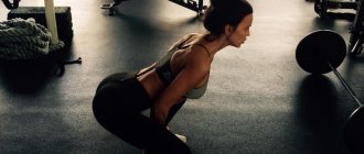
Exercises to train the gluteal muscles
From what was written above, we can notice that these muscles connect the pelvis and thigh. Thus, they become stabilizers for our legs. They also move the leg to the side. But the most important thing is the participation of the gluteus maximus muscle in hip extension. In order to develop them, you need to select exercises where these functions are present. There are several most effective ones:
- Sumo squats. All conditions are met here. Due to the wide stance of the legs, the leg abduction function is activated. When you come into a standing position from a sitting position, the hip extension occurs. And in order not to lose balance, the stabilizing function of these muscles comes into play.
- Romanian cravings. This exercise is aimed at working the buttocks. And it is one of the best for these purposes. The main function it involves is hip extension. Thus, it actively activates the gluteus maximus muscle. And by bending the legs at the knee joint, the biceps femoris muscle is excluded from the work.
- Leg abduction in crossover.
You can move your leg in different directions. Back, focusing on hip extension and working the gluteus maximus. And to the side, engaging the middle and small muscles.
Thigh muscles anterior group
It is this group that forms the thigh from the front. For men, the development of these muscles is of paramount importance. Several main muscles can be identified.
Sartorius
The shape resembles a narrow ribbon. And it is the longest muscle in our body. Its upper end originates from the anterior surface of the ilium. Then it descends obliquely down the front surface of the thigh. And is attached to the tuberosity of the tibia.
Functions:
Participates in flexion of the thigh and lower leg. With fixed hips, the pelvis tilts forward. It also participates in turning the hip inward.
Quadriceps femoris (quadriceps)
This is the largest muscle in our body. From the name it is clear that it consists of 4 heads:
Rectus muscle
The largest of the 4 heads. Located centrally on the front of the thigh. It is this muscle that forms the thickness of the leg. It originates from the lower surface of the ilium. Heading down, it passes into the common tendon. Which is attached to the tibial tuberosity.
Functions:
Due to the fact that the muscle crosses two joints: the hip and knee. She can not only straighten her leg at the knee. But also bend it at the hip.
Important:
When leaning forward during squats, this head is less involved in the work.
Vastus medialis
Located on the anteromedial (inner) surface of the lower thigh. From above it is slightly blocked by the rectus muscle. The shape resembles a drop of water. When well developed, it gives the thigh an expressive shape. From above, a thin tendon is attached to the inner side of the femur. Next, its fibers go down and pass into the broad tendon. With them it partially passes into the common tendon. And the remaining fibers are attached to the inner edge of the patella.
Functions:
Leg extension at the knee joint. Turn the shin inward. Stabilizes the knee, preventing it from falling inward.
Vastus lateralis muscle
Located on the outer part of the front surface of the thigh. Occupying almost the entire area. Slightly covered by the rectus muscle. This head gives shape to the thigh from the outside. Making your legs more massive. In bodybuilding, it is given special importance compared to other strength sports. It originates from the outside of the femur. Heading down, it passes into the common tendon of the 4 heads. And in some bundles it is woven into the lateral (outer) edge of the patella.
Functions:
Leg extension at the knee joint. Rotate the shin outward. Acts as a knee stabilizer. Not letting it fall out. That is, it performs the opposite action of the medial head.
Vastus intermedius muscle
Located on the front of the thigh, immediately under the rectus muscle. It is the weakest among all heads. The upper edge is attached to the anterior surface of the femur. And heading down it passes into the common tendon.
Functions:
Participates in leg extension at the knee joint.
Structure
So, in order to keep your legs in good condition, and if you want to pump them up, you need to know their structure and what each muscle group is responsible for.
So, let's look at what types of muscles our legs are made of:
1. Muscles of the back of the thigh.
2. The muscles of the front of the thigh (also quadriceps).
3. Calf muscles.
4. Buttock muscles.
5. Adductor muscles.
Also, each section consists of smaller muscles. Let's take a closer look.
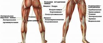
Posterior thigh
The most problematic area of the body for both sexes. In men, various imperfections appear here, and in women, cellulite is deposited. Therefore, it is this area that needs to be given the greatest attention.
Structure
- Semitransversalis muscle. It originates from the ischial tuberosity and is located on the back of the thigh. She is responsible for turning the tibia inward and moving the thigh itself
- Semitendinosus. It is also located on the lower thigh. The main purpose is to bend the leg at the knee.
- Biceps muscle. The second name is biceps, many people immediately think of the inflated part of the arm. However, in this case it is the hamstrings. Located in the posterior plane of the thigh. The role of flexion of the tibia at the knee.
Front thigh
- Quadriceps - quadriceps femoris muscle. The main purpose is to bend the knee. This name is due to the fact that the muscle itself consists of four more muscles. All quadriceps muscles are considered separate from each other, but they swing together at once.
Comprises:
- Rectus muscle. It has the longest length of the four, the muscle is located in front of the others and almost covers them.
- Vastus lateralis muscle. It is large in size and covers a huge area of the anterolateral part of the thigh.
- Intermediate wide. The most underdeveloped, located between the medial and lateral. Prevents joint bending when walking.
- Medial wide - occupies the anterior surface of the lower part of the thigh.
There are also adductor muscles; they guide the thigh itself.
Consist of:
- short;
- thin;
- large muscle.
Perform dynamic work in different sports.
Buttocks
The leg muscles originate from the gluteal muscles.
Consist of:
- Gluteus maximus muscle. Occupies almost the entire part of the outer thigh. It determines what your butt will look like. Its function is to tension the thigh and fix the body in a given state. There is an impact on the pelvis to hold the body above the femoral head.
- The gluteus medius muscle is a muscle of the outer group of pelvic muscles, located directly under the gluteus. It abducts the hip during contraction and also stabilizes the body during flexion and extension. Allows you to form the relief of the buttocks, so they need to be trained. The best exercise to help achieve results is squats. They need to be performed with some kind of load in order to achieve the best effect.
- The gluteus minimus is a muscle on the outer part of the pelvic muscles. The functions are the same as the middle one. Leg swings will help pump up this particular muscle.
Calf muscles
- Calf muscle. The triceps muscle, which is a two-joint muscle. Owns a large location. Formed by two heads of other muscles (medial and lateral)
- Soleus-shaped. It is located under the calf muscle and is smaller in size. Forms excellent leg relief. Takes part in the extension and flexion of the ankle joint and foot.
- Anterior tibial. It is located where the tibia ends, and accordingly it has this name. Participates in extension of the foot.
- Plantaris muscle. Small in size, but has a long tendon. Some people don't have it, but this is not abnormal for the human structure.
Exercises to train the front of the thigh
As you already understand, this muscle group extends the leg at the knee joint. The most effective exercises that fit these functions include:
- Front squats . Due to the fact that the bar is located in front, we will be forced to tilt it back a little. So as not to drop it. Therefore, most of the load will fall on the quadriceps. The main function involved in this exercise is leg extension.
- Bench press in a machine with narrow legs. Due to the narrow setting, the gluteal muscles are not included in the work. To shift the emphasis to the lateral head, place your feet together. On the medial side, place shoulder width apart.
- Stepping onto the platform. In this exercise, we focus on leg extension. And this is the main function of the quadriceps.
- Leg extension in a sitting machine.
It is a very dangerous exercise. Therefore, you should not use it on an ongoing basis. To avoid damaging your knee joints. From the name it is clear that it meets our requirements. Namely, it extends the leg at the knee joint. When we turn our toes to the side, we load the lateral head more. And inside, medial.
Muscle functionality
We don’t really think about how the muscles work during our movements; for us, all these actions happen automatically. The human body is a well-established mechanism, each element of which has its own functionality.
Quadriceps
Works with knee extension. Its constituent rectus muscle bends the body at the waist to an angle of 90 degrees, while it works separately from the other muscles.
Back of thigh
Basically, this muscle group works in the same way, namely: it extends at the hip joint and bends at the knees. Extends the body in the pelvic region, if the limb is fixed, and acts together with the gluteus maximus muscle. When the knee is bent, it allows for inward and outward rotation of the lower leg.
Buttocks
They function by bringing the hips together, and also perform rotational movements in and out. If the limbs are fixed, then the body is extended and the pelvis is tilted to the sides.
Adductors
They are also called adductors or “moral muscles.” In addition to their key role in hip adduction, they are involved in pelvic flexion and extension and axial rotation of the limb. Another important feature is that they help stabilize the body. The training of these muscles helps to quickly maneuver, which is especially important during active sports.
Posterior surface of the leg
In addition to bending the knee and foot, the muscles here twist the lower leg, supinate the foot, bend the toes, and press them toward the ground while standing.
Medial thigh group: adductors
This area is very problematic. And most athletes are developmentally delayed. Because they are not given due attention during training. And without their development, the thigh will not look aesthetically pleasing. Large and developed from the outside. And atrophied from the inside. This group includes the following muscles:
Thin muscle
It has the most superficial location, on the inside of the thigh directly under the skin. The upper edge is attached to the anterior surface of the pubic bone. Then it goes down, around the medial epicondyle of the femur. And is attached to the tibial tuberosity.
Functions:
Takes part in hip adduction and flexion. It also rotates the lower leg inward and takes part in its flexion.
Adductor longus muscle
This is a flat muscle, vaguely shaped like a triangle. Located slightly in front, closer to the quadriceps. It originates from the pubic bone. As it moves downwards, it widens and attaches to the middle part of the femur.
Functions:
Hip adduction, flexion and internal and external rotation.
Adductor brevis muscle
It also has a triangular shape. It is located slightly deeper than the adductus longus. Starts from the front surface of the pubic bone. Next it goes down and is attached just above the middle of the inner surface of the femur.
Functions:
Involved in hip flexion and hip retraction. Stabilizes the pelvis, preventing it from tilting back.
Adductor magnus muscle
This is a wide and thick muscle. She is the strongest of the adducting group. It is located deeper than the long and short muscles, on the inside of the thigh. It originates from a powerful tendon from the bottom of the pubis and ischium. Heading down, it spreads out like a fan onto the wide tendon. And it is attached to the entire inner surface of the femur, and partly to the medial epicondyle.
Functions:
Adducts the hip. Takes part in its bending. Participates in stabilizing the pelvis.
Bones and joints
The first section is involved in the construction of the pelvic joint (it includes the pubic, ilium and ischium bones, the sacrum and the thigh muscles, which serve for strengthening and normal functioning; the underlying elements are attached through the hip joint).
The second involves the femur. It is the largest in the body. The description is similar to a tube, bent at a certain angle, inside which is yellow bone marrow. Tendons and muscles are attached to its body to provide mobility to the leg; the lower part takes part in creating the knee joint.
The third is formed by the tibia and fibula. The first is part of the knee joint and has condyles to which tendons are attached. The second is placed lower and serves to strengthen the knee.
The foot performs the function of supporting the surface and consists of a tarsus, metatarsus and phalanges of the fingers (the anatomical structure is represented by diagrams that include a combination of three bones, and the first toe - two).
Exercises to train the adductor muscles
As you can see, these muscles are responsible for a stable position. Don't let your pelvis fall back. And of course they bring their legs towards each other. Their development will help improve your posture. Strengthen your inner thighs. Due to this, we can increase strength in many basic exercises. The following are good for their development:
- Plie squats. This is a great basic exercise. Which meets all anatomical functions. By placing the legs wide and turning the feet, we stretch the adductor muscles as much as possible. Also, when coming out of a squat, the thighs are brought towards each other.
- Leg abduction in the simulator. From the name it is clear that the exercise meets our requirements. But when performing it, you must follow the correct technique. There should be no jerky movements. To avoid injuring the adductor muscles.
- Adduction of the leg in a crossover
. Also a common exercise that allows you to work the adductor muscles.
Basic exercises for all leg muscle groups with names and photos
Front Squats
For some reason, recently this species has been unreasonably forgotten. And very much in vain.
First of all, it should be said that it is performed with a barbell on the chest. It is extremely simple to perform and at the same time removes some of the load from the back.
Lunges
They definitely need to be included in the training program. They work the muscles well and are also the least traumatic.
In the first stages, practice in front of a mirror. The feet should be clearly under the hips to create a straight line. As you inhale, take a step forward. The weight should be evenly distributed. Three right angles should form: between the quadriceps and the torso, in the right and left knee. Then - to the starting position.
Simple enough isn't it?
Thigh muscles posterior group
These muscles are antagonists for the anterior group. That is, they perform opposite functions. The front one extends the leg, and the back one bends it. In their training, the main thing is to exclude the gluteal muscles from the work as much as possible. If we don't do this then they will take on most of the load. This group includes:
Biceps muscle (biceps femoris)
Located along the outer (lateral) edge of the back of the thigh. This muscle is the main one in this group. And most of the exercises are aimed at its development. Consists of two heads:
- Long. Which starts from the ischial tuberosity, with a small flat tendon.
- Short. It originates from the inner surface of the lower half of the femur.
Below, these two heads unite into one powerful abdomen. Which passes into a narrow tendon. And bending around the back of the lateral epicondyle of the femur, it is attached to the head of the fibula.
Functions:
Extends the thigh together with the gluteal muscle. Bending the leg at the knee joint. And in this position it can turn it outward.
Semitendinosus muscle
This is a long and thin muscle. It is located closer to the inner (medial) edge of the back of the thigh. Starts from the ischial tuberosity. Heading down, it passes into a thin tendon that wraps around the medial epicondyle of the femur from behind. And is attached to the tuberosity of the tibia.
Functions:
Participates in hip extension and leg flexion at the knee joint.
Semimembranosus muscle
Located along the inner edge of the back of the thigh. Covered below by the semitendinosus muscle. It originates from the ischial tuberosity. Heading down, it goes around the epicondyle of the femur and attaches to the medial condyle of the tibia.
Functions:
Extends the hip. Bends his leg. It also turns the shin inward with the knee joint bent.
Hamstring workout
That is, as we see, exercises should perform two main functions. This is hip extension or leg flexion. We can also use foot rotation in some exercises. When bringing your toes together, the semitransverse muscle is loaded more. When extended to the sides, hamstrings.
- Deadlift on straight legs . This is a basic exercise. Which engages the hip extension function when coming out of a bend. Thus, all the muscles of the posterior group are activated. And since the legs remain straight when tilted. Most of the load will be taken by the back of the thigh.
- Squats with wide legs. Also applies to basic exercises. Involves two functions. Bend the leg at the moment of the squat itself. And hip extension when returning to the starting position. Squats also work all 3 gluteal muscles. And the adductor muscles receive a small share of the load.
- Leg bending in the simulator. Here I think everything is clear. The exercise gives us the opportunity to work the back of the thigh in isolation. By bending the leg at the knee joint.
- Taking the leg back in a crossover. This exercise is mainly performed by girls. It allows you to work all the muscles of the posterior group in isolation.
Hip
This part of the body has the shape of a truncated cone - its apex is the knee, and its base smoothly borders the body. This appearance is due to the structure of the soft tissues - the upper segment of the thigh contains a large number of muscles. In the lower section, the muscles smoothly transform into wide and strong ligaments, as a result of which the volume of the limb decreases.
The thigh, as a part of the body, has clear boundaries, although an ordinary person is unlikely to be able to correctly indicate them. Therefore, we should consider exactly how it is located in relation to the torso and lower leg:
- The upper border is not transverse throughout - in front it runs along the skin inguinal folds, going obliquely downwards. From the side, the leg is delimited from the body along a line drawn through the iliac crest. From behind, the border acquires a transverse direction, passing in the gluteal fold. Its general internal direction corresponds to the plane drawn through the hip joint.
- The lower border of the thigh does not have such structural features, and is calculated quite simply - in relation to the patella. The upper pole of the patella is determined, after which a perpendicular line is drawn 5 centimeters above it.
Knowing the correct boundaries of any part of the body allows the doctor to accurately assess the localization of pathological processes, and also helps to easily find large vessels or nerves in their projection.
Skeleton
The entire static and functional load in this part of the body is assumed by a single bone - the femur. It is the largest indivisible structure of the musculoskeletal system in all respects - size and weight. According to the anatomical classification, the femur has a tubular structure, which is characteristic of the most loaded and durable formations in the skeleton.
Since it is only one supporting element of the upper segment of the leg, it has to take on the interaction with all the soft tissues. Therefore, the femur has a rather interesting structure:
- The upper part consists of the head and neck, which are part of the hip joint. In relation to the segments lying below, they are located at a slight angle. This device not only provides good support, but also increases the range of motion in the joint.
- Further, the neck passes into a large tuberous formation - the greater and lesser trochanter of the femur. They are the attachment point for the large gluteal muscles.
- Then the largest and longest segment begins - the body of the bone. It has a characteristic tubular structure, expanding slightly in the lower section. On its posterior surface there is a rough line - a fixation area for some thigh muscles.
- The lower section consists of rounded extensions - it is divided transversely by a wide depression. These parts are called condyles - they are normally covered with articular cartilage and form the upper half of the knee joint.
The head and neck of the femur have a relatively isolated blood supply, which affects the rate of healing when they are damaged.
Soft fabrics
Between the skin with fatty tissue and the muscle tissue of the upper leg there is another large formation - the fascia lata of the thigh. It is a large sheath of connective tissue that collects all the muscles of the anterior and lateral sections into one large bundle. The durable outer shell gives them the support they need, allowing them to work more efficiently and smoothly.
Inside the muscle bundles there are also tendon septa, dividing them into three groups. Each of them performs a certain range of movements during contraction:
- The anterior group consists of two long and strong muscles - the sartorius and the quadriceps femoris. Their purpose is to flex the leg at the hip joint, as well as extend the knee. The quadriceps muscle in the lower part forms a powerful and wide tendon, which passes through the kneecap to the lower leg.
- The posterior group is formed by thin and long muscles - biceps, semimembranosus and semitendinosus. On the contrary, they carry out extension at the hip joint and flexion at the knee joint. And with fixed legs, their contraction allows you to return the torso from a tilted position.
- The internal group consists of small short muscles - the pectineus and gracilis muscles, as well as the adductor magnus, brevis and longus. As a result of their coordinated work, the hip is adducted and rotated outward.
The peculiarity of the thigh muscles is their dual purpose - they take on both powerful static and dynamic loads, often combined with each other.
Vessels and nerves
The vast majority of these formations are located in the space located between the anterior and internal muscle groups. Starting from the upper border, the main vascular bundle passes there, providing blood supply to the entire lower limb. The nerves are divided according to the opposite principle - the largest of them, on the contrary, runs in the back of the thigh.
In general, the arrangement of blood vessels and nerve bundles is of the main type, characteristic of such a large segment of the limb. Therefore, they should be considered within these highways:
- Arterial vessels are represented by the large femoral artery, which passes to the limb from the pelvic cavity. It runs in the intermuscular cavity along the inner surface of the thigh, giving off a deep branch to supply almost all of the muscles listed above. The main trunk, just above the knee, goes deep into the soft tissues, penetrating the popliteal fossa and extending to the lower leg.
- The venous system consists of two parts - the femoral vein represents its deep part, and the large saphenous vein is a superficial vessel. Just below the inguinal fold they merge, forming a common vein that extends into the pelvic cavity.
- The innervation of the thigh is provided by two systems of nerves located on its opposite sides. Together with the vessels, the femoral nerve emerges on the inner surface. The most powerful similar structure in the body, the sciatic nerve, runs behind it.
The main type of blood supply and innervation makes the legs vulnerable to injury, since if a vessel or nerve is damaged at the hip level, the entire limb suffers.
Calf muscles
These muscles are stabilizers for our feet. And is involved in bending the leg at the knee joint. The muscles of the anterior group are responsible for turning the foot, flexing and extending the fingers. But as I said earlier, we will not consider them. We are more interested in the back group. Which includes:
Triceps surae muscle
This is the main muscle that can be trained, and we can influence its shape.
Consists of two muscles:
Calf
lies on the surface and has two powerful heads.
- Medial. It is located closer to the inner part of the back surface of the lower leg. And it is more powerful. Starts from the posterior surface of the medial epicondyle of the femur.
- Lateral. This head, on the contrary, is located closer to the outer part of the lower leg. And it starts from the posterior surface of the lateral epicondyle of the femur.
These two heads are directed downwards and in the middle of the lower leg they are connected into one powerful tendon. The second muscle is the soleus.
This is a flat muscle. Located under the calf. One edge is attached to the head of the fibula. And the other to the inner surface of the tibia. And going down, it connects to the tendon of the gastrocnemius muscle. Which attaches to the heel bone. This tendon is known to many as the Achilles tendon.
Functions:
Participates in bending the leg at the knee joint. Flexes the foot. It is also a stabilizer for the lower leg muscles.
Plantaris muscle
This is a very short muscle that has a long and thin tendon. It originates from the lateral condyle of the femur. It goes down and passes into a narrow tendon that lies between the soleus and gastrocnemius muscles. Attaches to the heel bone.
Functions:
Helps bend the leg at the knee joint. Participates in raising the foot. Tenses the capsule of the knee joint.
Anatomical structure of the human leg
Functions
The leg has many functions:
- walking;
- run;
- jumping;
- crawl;
- swimming;
- support, etc.
Parts
If you remember the anatomy, the leg has three parts - thigh, lower leg, foot.
Hip
Performs a protective function. It consists of the femur, the patella, and on top it is covered with the quadriceps, biceps femoris and flexor muscles.
Shin
It has a fairly simple structure and consists of two bones of different lengths, called the fibula and the tibia.
The latter connects the tibia and femur at the knee joint and is the second largest in the human leg.
Foot
Formed from many small bones. The foot or sole is the point of contact with the surface of the earth. And the opposite side has a name - the back.
The foot is divided into 3 sections:
- the anterior one, consisting of the toes and balls of the foot;
- middle - arch of the foot. The concept of arch includes that part of the foot where it does not reach the ground;
- back - heel.
The foot is much more complex in structure and has more than 26 bones and 33 joints. The structure of the foot and hand are very similar, differing only in the degree of load tolerated. The muscles and bones of the foot are many times stronger, but they cannot boast of the mobility of the hand.
Leg areas
The leg consists of the following areas:
- anterior + posterior thigh areas;
- anterior + posterior knee areas;
- anterior + posterior areas of the lower leg;
- anterior + posterior, outer + inner parts of the ankle joint;
- back of the foot;
- sole.
Ankle
The largest bone is the talus. At the top there is a block with a protrusion, connected by the tibia and fibula.
On the side there are projections of bone called ankles. On each surface of the joint there is hyaline cartilage, which performs shock-absorbing and nutritional functions.
The joint itself is complex in structure, as it consists of more than two bones. It has a block-like shape.
Ligaments
The ankle ligaments play a huge role. They are the ones who limit movement in the joint, protect it, and hold the bone structures together.
In general there are three groups:
performs a connecting function between the bones of the lower leg . It includes the following ligaments: - lower, preventing internal rotation of the bone; - lower anterior fibular, which prevents the foot from turning to the outside; - interosseous; - transverse, fixing the foot.- deltoid ligament , representing the outer lateral fibular fibers that strengthen the outer edge. These are: - heel; - anterior talar; - posterior talus.
- preventing bones from shifting . This group originates on the medial malleolus and consists of: - tibial calcaneal ligament; - tibial scaphoid; - front ram; - rear ram.
Calf muscles
The lower leg consists of 20 muscles responsible for raising, lowering, and moving the leg and toes. A large number of muscles begin at the back of the knee and end at the foot. They are the ones who set the leg in motion. Each muscle has its own purpose and function.
The lower leg has three muscle groups:
- anterior, responsible for extension of the feet and fingers;
- external, driving the outer edge of the foot;
- back, allowing movement of the foot and toes.
The strongest muscle is the gastrocnemius. Its origin is located at the heel bone of the foot, on which it is held by the calcaneal tendon.
The calves are made up of two muscles: the gastrocnemius and the soleus. The gastrocnemius is a large, knobby muscle formed from two parts that form a diamond shape. The second, soleus, is completely flat and hidden by the gastrocnemius.
When you walk, run, or otherwise move your legs, the calf muscle pulls up your heel, which is what makes your legs move.
An important part is the Achilles tendon, which gives rise to three muscles at once - the gastrocnemius, plantaris and soleus. It is thanks to this tendon that a person can run, jump, walk and move. Often this is the part that is subject to stretching and tearing.
Functionality
The lower leg is designed to provide the necessary mobility when walking. The muscles of the joint work harmoniously and perform extension, flexion, rotation of the ankle, and also create shock absorption.
Blood supply
The junction of the tibia and fibula is located below the knee joint.
The lower leg is fed through the tibial arteries - posterior and anterior, starting under the knee.
The arteries branch and surround the joint on all sides.
Veins run next to the arteries. Blood circulates through the internal and external networks, forming the tibial and saphenous veins.
Calf muscle training
Based on what was written above, we can draw a conclusion. That this muscle group receives part of the load in almost any exercise for the legs where we bend them at the knee joint. Plus, they act as a stabilizer for the foot and are also used when rising onto the toes. There are several specialized exercises:
- Calf raises in a special machine. There are a lot of such simulators. There is an option where we perform the exercise standing, sitting and even lying at an angle. Therefore, choose any exercise machine that is in your gym and start raising your toes.
- "Donkey." Refers to a number of rarely seen exercises in modern gyms. But by bodybuilders of the golden era, it was in demand. By tilting the body forward, we stretch the buttocks and the back of the thigh. Therefore, they will not be able to turn on and take the load from the calf muscles.
- Calf raises with dumbbells. This is similar to the first exercise, but instead of a machine, a dumbbell weight is used. For greater stability, take it in one of the hands and perform lifts on each leg in turn.
As you can see, anatomy is an interesting science and its knowledge is very useful. Especially for people who want to develop their body. You don't need to know all the scientific terms. The main thing is to understand what the muscle is roughly attached to. To the pelvis, to the thigh or lower leg and what function it performs. Then you can choose the right exercises.
Good luck to everyone in your training!