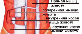
Abdominal muscle structure:
We humans are all the same. Our muscle set is the same as that of any other person. This also applies to the abdominal cavity. These muscles are responsible for the following functions:
- Form the abdominal wall
- Protects and keeps internal organs in a stable position
- Support our body and form correct posture
Abdominal muscles:
- Rectus muscle
- Oblique (external and internal)
- Transversus muscle
It is conventionally accepted to divide these abdominal muscles into special groups, such as the anterior, lateral and posterior walls of the abdomen.
What is the transverse abdominis muscle?
The transverse abdominis muscle is one of the main muscles that makes up the bulk of the weight. It was named because of its structure; it is also called “skeletal”. Located in the bones of the skeleton, when contracted, it causes the entire torso and joints to move. It is also involved in the functioning of the transverse spinalis muscle. The muscle runs along the spine, represents the middle back muscle, and is actively involved in abdominal training exercises. The permanent membrane is a thin muscle-tendon plate, the muscle bundles of which are directed transversely. They are located deep in the abdomen, they cannot be trained to create relief, but they are responsible for the tone of the abdominal cavity.
Rectus abdominis muscle:
So, this muscle, by its specificity, is flat and long, consists of special, thin bundles of muscles that are located vertically. It is this muscle that those who dream of expressive abs love so much. It starts from the chest, runs along the entire abdomen, and is attached at the end to the pelvic bone. A special layer of connective tissue, or the linea alba, divides the rectus muscle into two parts.
Main function:
- Twisting of the body in the lumbar spine
- Raising the pelvis with a fixed chest
- Increased intra-abdominal pressure
- Lowering the ribs, exhale
The rectus abdominis muscle is endowed with great capabilities in terms of lifting force and cross-section. It acts as a powerful flexor of our spine. It is worth noting that in the process of fixing the chest and during its contraction, the pelvis rises, and not lowers the chest in its direction.
The rectus muscle has an interesting feature - it can contract in separate areas, and not all at once. Because of this condition, you can focus your training program on individual areas of this muscle, selecting the necessary exercises.
Exercises to develop abdominal muscles
To keep your abs in order, you need to perform various exercises to maintain the tone of their muscles. Their implementation is a preventive measure for the human respiratory system, to maintain the kidneys and spleen in the correct position, to prevent hernias and protrusions.
For women during the postpartum period, it is important to strengthen the abdominal muscles. It must be remembered that it is necessary to start with simple complexes so as not to create a hernia. And the intensity can be increased six to eight months after the baby is born.
All people need information that during intense sets of exercises, the abdominal muscles begin to ache. This is directly related to the irregularity of performing complexes or, conversely, to too much stress on the muscles. To avoid painful sensations, you need to gradually increase the load.
Sometimes there are times when pain in the abdominal muscles is not associated with exercise, but is associated with heavy physical work. In such cases, a hot bath or sauna is indicated.
Let's name the most popular and effective exercises for the abdominal muscles.
Raising the body from a lying position on a horizontal surface
Raising the torso effectively affects the abdominal muscles. This complex requires strict adherence to the rules for its implementation. It is imperative to perform the movements in full. It is necessary to lie on a flat, hard surface, bend your lower limbs at the knees, and firmly fix your feet.
Hanging leg raises on the bar
Hanging leg raises have a beneficial effect on the abdominal muscles. When performing this complex, you need to hang on the crossbar, then raise your lower limbs up, then lower them. It is important to remember that it is not advisable to bend your arms and raise your pelvic bones as little as possible. You should not swing the body and after each completed complex, be sure to wait until the body takes a strictly vertical position.
Raising outstretched legs
Raising your legs in an extended position has a qualitative effect on the abdominal muscles. You need to lie on the floor and place your hands, palms down, under your buttocks. Raise your legs in an extended position almost vertically as much as you can. Next, the lower limbs must be returned to their original position, and, trying not to touch the floor with your feet, repeat the entire complex from the beginning.
External oblique muscle:
The peculiarity of this muscle is that it is considered the widest on the surface of our body and is located on both sides of our torso. Its fibers are arranged in such a way that they run from top to bottom and medially, to the midline of the body. It originates from the surface of the sternum on the side, or more precisely, from the 8 lower ribs. It is clearly depicted below.
Main function:
- Helps rotate our body in opposite directions (subject to unilateral contraction)
- Provides downward traction of the ribs, as well as flexion of the torso (subject to bilateral contraction)
- Helps with lifting and carrying various weights
- Keeps our body upright
What functions do the abdominal muscles perform?
The transverse abdominal muscles form the deepest, third layer of the abdominal wall. The muscle bundles are laid horizontally. They bend back at the waist. When muscles contract, the abdominal cavity decreases, the stomach tightens, and the ribs tighten. Their parallel work promotes bending of the torso forward and in different directions. They are responsible for stabilizing the lumbar and pelvic bones and act as a corset in the abdominal area.
Location and structure of the main oblique muscles
External and internal oblique abdominal muscles
There are two oblique abdominal muscles - external and internal. They are similar in size and slope, but differ in attachment areas and location. The functions of these elements are not interchangeable, but complement each other.
The oblique muscles are paired elements of the body, so all exercises for their development are performed first on the left, then on the right side.
Features of the external oblique abdominal muscle
External oblique muscle
This muscle originates from the outer surface of the 5th-12th ribs and is attached in several places: to the linea alba at the very top, to the outer lip of the iliac crest and gradually passes into the groin area. In the pelvic area, the external oblique muscle attaches to the inguinal ligament, then extends to the pubic tubercle and crest.
Innervation of the external muscle occurs through several groups of nerves:
- ilioinguinal in the lower part;
- iliohypogastric in the middle region;
- intercostal - in the place where the muscle originates.
Among the structural features of the external tissue, one can highlight the fact that in the area of attachment to the ribs it is tightly intertwined with the serratus anterior muscle, as well as with the latissimus dorsi muscle in the area of departure from the lowest ribs.
One of the most important functions of this element is the formation of the rectus abdominis muscle together with the internal oblique fascia (formation of the vaginal leaf). The external oblique muscle has many primary functions:
- participates in the breathing process - at the moment of exhalation it is responsible for lowering the ribs;
- supports the position of the abdominal organs, partially the chest and pelvis;
- ensures normal intra-abdominal pressure;
- responsible for the opposite rotation of the chest and flexion of the spine without injury;
- directly involved in physiological processes associated with other muscles: waste elimination, coughing, childbirth;
- responsible for correct posture;
- protects the spine from injury under any load.
The externus muscle is a relatively thin layer of tissue covering a large area of the abdominal area. Any damage can cause severe pain and leave a person unable to move.
Features of the internal oblique muscle
The internal muscle refers to the main abdominal muscles. It is located in the anterior zone of the body. It lies immediately below the externus muscle, but above the transverse abdominis muscle, and runs lateral to the rectus muscle.
The internal oblique abdominal muscle is one of the most powerful and strong, and has high plasticity with the appropriate level of training. Borders on the lumbar region. The oblique muscle extends obliquely from the aponeurosis of the rectus muscle:
- attached to the linea alba;
- connects to the pubis;
- extends from the outer surfaces of the 4 lower costal arches.
In combination with the extrinsic muscle, it provides slight lateral bending. Like the external muscle, it has a paired structure - on the left and on the right. The main functions of this element include:
- ensuring tilt of the pelvic area;
- bilateral contraction of the pelvic region;
- flexion of the thoracic and lumbar spine;
- tilt and rotation of the thoracic and lumbar region towards the contracted muscle;
- forced exhalation.
Innervation of muscle tissue occurs by intercostal nerves from Th-6 to Th-12, in the middle zone - by the iliohypogastric nerves, and in the lower zone - by the ilioinguinal nerve trunk. The upper, lower and intercostal arteries located in the posterior space, as well as the muscular-diaphragmatic artery, are responsible for the blood supply to the element.
The internal oblique muscle of the abdomen is an indispensable element of movement and breathing, when damaged, all functions of the body responsible for its life support are partially disrupted.
Transverse abdominis muscle
Functions of the transverse muscle
It is not included in the group of oblique fibers, but has the tightest connection with them. This muscle is located very deep relative to the anterior lateral abdominal wall; the internal oblique muscles overlap it. The main function is to stabilize the lumbar spine.
The transverse muscle originates from the inner part of the 6 lower ribs and is attached to the transverse processes of the lumbar vertebrae, the iliac crests and the thoracolumbar fascia. Additionally connects to the white line of the abdomen. The main functions include: forced exhalation, reducing the size of the abdominal cavity.
Muscles that form the anterior abdominal wall
1. Rectus abdominis (musculus rectus abdominis). Things didn’t work out with high-quality cadaveric material at my university, so I saw this muscle for the first time during an operation (it was my third year, I think). It looks like a fairly wide strap that stretches strictly vertically across the entire stomach.
More precisely, two wide straps, because the muscle is steamy.
The rectus abdominis muscle is divided by tendon bridges (intersectiones tendinae), which are located across the fibers of the muscle. You can see the white cross stripes in both illustrations. It is these jumpers that form the “cubes” that are clearly visible in athletic people.
The rectus abdominis muscle is enclosed in the vagina (vagina musculi recti abdominis), which consists of aponeuroses of the muscles of the lateral abdominal wall.
Origin: pubic bone, the space between the pubic syphysis and the pubic tubercle.
Insertion: anterior surface of the xiphoid process of the sternum; outer surface of the cartilage of 5-7 (inclusive) ribs.
Function: bends the spine, with the chest fixed (for example, hanging on a horizontal bar), raises the pelvis.
2. Pyramidal muscle (musculus pyramidalis)
This is a rather small muscle, it is not always possible to see it. To see it, you need to look for a small, triangular-shaped muscle in the pubic area. In the figure above, in which we noted the rectus abdominis muscle, we can also find the pyramidal muscle:
Origin: pubic bone, anterior to the beginning of the rectus abdominis muscle;
Attachment: lower part of the white line of the abdomen;
Function: tightens the linea alba.
Feature: the muscle is vestigial. According to Sapin's textbook, the pyramidalis muscle may be completely absent.
Lateral intertransverse lumbar muscles[edit | edit code]
Lateral intertransverse lumbar muscles
Lateral tract, intertransverse system
Lateral intertransverse lumbar muscles
(
mm. intertransversarii lumborum laterales
) straighten the spine during bilateral contraction and tilt it to the same side during unilateral contraction. Like the medial intertransverse muscles, they stabilize the lumbar spine and keep the vertebrae from moving laterally.
Home[edit | edit code]
- Costal processes of all lumbar vertebrae
- Transverse process of T12 vertebra
Attachment[edit | edit code]
- Costal processes of vertebrae L5-L1
- Transverse process of T11 vertebra
- Iliac tuberosity
Innervation[edit | edit code]
- Anterior rami of spinal nerves T12-L5
Features[edit | edit code]
These muscles are formed from the ventral germ layer and are therefore innervated by the anterior (ventral) branches of the spinal nerves
Functions[edit | edit code]
| Synergists | Antagonists |
| Intervertebral joints and discs (lumbar spine) | |
| Extension (bilateral contraction) | |
| mm. intertransversarii lumborum mediales All deep back muscles of this area | m. rectus abdominis m. obliquus externus abdominis m. obliquus internus abdominis |
| Tilt in the same direction | |
| mm. intertransversarii lumborum medialis All deep back muscles of this area (without spinous and interspinous muscles) m. rectus abdominis m. obliquus externus abdominis m. obliquus internus abdominis m. quadratus lumborum | All deep muscles that act as synergists on the same side become antagonists when contracted on the opposite side |
