Anatomy
The musculus triceps brachii consists of several main structures. They represent three muscle bundles (heads), which is reflected, in fact, in the name. The triceps brachii muscle extends all the way across the back of the humerus.
The three heads of the muscle have their own names, reflecting their structural location: lateral, long and medial.
- The first of these (caput laterale) is attached muscularly and tendon to the surface of the humerus (outside).
- The middle position is occupied by the caput longum, it is the longest and is attached to the scapula (subarticular tubercle).
- The third is caput mediale, attached one third below the head of the humerus, at the back, and has a fleshy upper part.
All three of these heads are combined into one muscle, which below passes into a tendon formation attached to the ulna.
Structure
The human shoulder consists of bones (scapula, clavicle, humerus), ligaments (coracoid or acromial, superior or transverse), joint, as well as soft and connective tissues. All of them are parts of the free upper limb, adjacent to the group of joints of the upper body (the shoulder girdle). The fabrics hold our arms in the shoulder joints and provide them with freedom of movement.
In women and children, the shape of the shoulder is smoother - cylindrical. This is due to the distribution of the fat layer. In men, this part of the limb looks larger and more prominent. With a sufficient amount of physical activity and a certain diet, the relief of the shoulder muscles looks more pronounced. This is due to the fact that the large biceps and triceps muscles are located not deep, but just under the skin.
It should be noted that the muscles and individual bundles of muscle fibers of other parts associated with it are responsible for some movements of the shoulder. For example, trapezius, deltoid, latissimus dorsi and others. The deltoid muscle gives the shoulder joint its roundness.
Important! Many athletes call all the muscles of the shoulder girdle, as well as the muscle fibers of the chest and neck, shoulders, but from an anatomical point of view this is incorrect. Experts in human anatomy classify as the shoulder musculature only the tissue of the free limb from the shoulder joint towards the elbow.
The muscle tissues of this part of the body are divided according to topographical characteristics relative to the humerus into anterior and posterior, as well as functionally into flexors and extensors. The anterior group of shoulder muscles is responsible for flexion, and the posterior group is responsible for extension.
The structure of the flexors and extensors allows the arms to move in all planes: abduction, adduction, flexion, rotation.
The posterior and anterior groups are separated from each other by intermuscular partitions.
This group includes the following muscle tissues:
- two-headed,
- shoulder,
- coracobrachial (acromiobrachial).
The brachial muscle tissue lies directly under the biceps, but is deeper. Attaches to the upper part of the humerus and to the tuberous surface of the ulna.
The coracobrachialis has an elongated, pointed shape, which is why it got its name. It originates from the coracoid process of the scapula, and the other end is attached to the inside of the shoulder bone.
The posterior group of muscle tissues includes:
- triceps (triceps),
- elbow muscle,
- ulna.
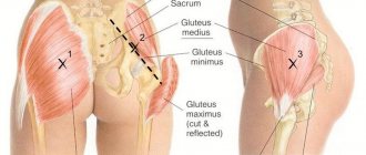
The triceps (triceps) has three attachment points - three heads: long, lateral and median. The beginning of the long head falls on the humeral process of the scapula, and just below the muscle passes into a common tendon with the other heads. The lateral and median heads are adjacent to the posterior surface of the humerus bone. As a result of the connection of the three heads, a rather prominent muscle is formed, which passes into the flat wide tendon of the triceps brachii muscle and is attached to the olecranon process of the ulna.
The muscle tissue of the elbow joint prevents pinching of the elbow joint.
Note. This muscle tissue is considered a non-permanent anatomical formation. Experts classify it as part of the triceps muscle.
The ulna is a continuation of the median head of the triceps. The fibers originate from the lateral epicondyle of the humerus and attach to the posterior surface of the olecranon.
A pyramid-shaped cavity is visible on the inside of the shoulder joint. This is the armpit. It has three walls formed by the muscles of the chest, back and shoulder girdle. Vessels and nerves pass through it, providing the function of the upper limb.
Triceps work
The function of the triceps muscle, or rather, the most important of them, is extension of the forearm. This is the same hand movement when, when the elbow is extended, the entire arm straightens. But its medial part is responsible for this. The main antagonist is the biceps muscle.
The long head of the biceps is responsible for shoulder extension (adduction to the body). The antagonists for it are the deltoid, pectoral and part of the biceps. The medial head is usually not easy to palpate, since it lies deeper, under the long head.
It is the biceps that acts as a counterweight to the triceps when flexing and extending the arm at the elbow joint. This is worth remembering when pumping muscles in the gym. Some athletes have “bloated” arms that look funny when the triceps are dominant.
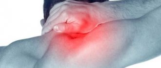
Diagnostics
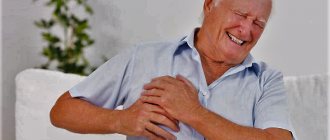
Problems with CPM are manifested by severe pain, so patients do not tolerate discomfort for long and consult a doctor with complaints of discomfort in the anterior deltoid area. When palpating the tissues, the doctor looks for painful lumps that indicate the presence of pathology. By inserting your fingers deeply under the armpits, you can feel the back of the muscle complex and tendon.
Symptoms of shoulder joint diseases
By pressing and holding their fingers, doctors feel the pulsation of the artery located near the coracobrachialis muscle. To palpate the muscle itself, the bundle of nerves and blood vessels needs to be moved slightly.
Diagnosis includes assessment of function:
- range of motion – if the CM is damaged, movements are limited and are accompanied by pain when flexing and extending the shoulder;
- muscle strength - examined in a sitting position, with the shoulder abducted to forty-five degrees, the arm partially bent, and the hand pronated. In this position, the doctor presses on the elbow, pulling down and pressing from behind. If the muscular strength is weakened, the patient's arm will be pliable and will not resist;
- back rubs are a typical test that doctors ask patients to do. A sick person sits and puts his hand behind his back, trying to rub his back with the back of his hand. When muscle tissue is damaged, the patient cannot perform movements in full and only reaches part of the back, and due to pain and discomfort, cannot rub it.
Triceps in sports
The triceps brachii muscle, whose functions are varied, can be the most important in some sports, where extension of the forearm during dynamic loading is of paramount importance. Here are some examples:
- javelin-throwing;
- shot put;
- volleyball, handball, basketball;
- boxing;
- fencing and other sports.
Also, in some cases, the triceps performs the task of “holding the load,” for example:
- cycling;
- archery;
- Weightlifting.
As a muscle that performs active shoulder extension (and adduction to the torso), the triceps is important in the following sports:
- rowing;
- swimming (all types);
- kayaking.
Participation in sports[edit | edit code]
The coracobrachialis muscle is involved in shoulder flexion and performs dynamic work during breaststroke and backstroke swimming, bowling, boxing, and static work when stabilizing the upper limb in front during boxing or in an elevated position in gymnastics. As a shoulder adductor, it performs dynamic work in breaststroke swimming and hockey, and both dynamic and static work in ring gymnastics. Participates in exercises of all sports related to movements in the forward arm (hockey, discus throwing, shot put, tennis). In addition, it prevents caudal displacement of the humeral head when lifting heavy objects.
| Kind of sport | Movement/hold | Function | Load | Types of abbreviations |
| Swimming | Breaststroke and backstroke - carry phase | Shoulder flexion | Strength endurance | Dynamic concentric |
| Breaststroke | Shoulder adduction | Strength endurance | Dynamic concentric | |
| Boxing | Hit from bottom to top | Shoulder flexion | Fast, explosive | Dynamic concentric |
| Main hand hold in front | Stabilization of the raised upper limb from the front | Strength endurance | Static | |
| Gymnastics | Handstand; with all elements requiring elevation of the upper limb; torso hold | Stabilization of the upper limb in an elevated position | Strength endurance | Static |
| Hockey | Hit with a stick | Shoulder adduction | Fast, explosive | Dynamic concentric |
| Discus throw | Throw | Anteversion from a raised position | Fast, explosive | Dynamic concentric |
| Tennis | Forehand | Anteversion from a raised position | Fast, explosive | Dynamic concentric |
| Weightlifting | Thrust phase | Stabilization of the humeral head in the glenoid cavity | Fast | Dynamic concentric |
A question of aesthetics
Many people ask: “Why train triceps?” From the above material it becomes clear that this, first of all, needs to be done by athletes. Do ordinary people need to “pump up” this muscle?
The answer to this question lies in a person’s desire to work on himself. The shape of the forearm mainly depends on the condition of the triceps muscle of the arm.
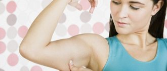
Sagging skin often occurs in women who are overweight or who have lost weight dramatically. By “tightening” your triceps, you can aesthetically improve the appearance of your shoulders and start wearing beautiful short-sleeved dresses.
Functions of the muscles of the shoulder girdle
Table describing the main functions:
| Name | Start | Fastening | Function |
| Deltoid | Acromial end of the clavicle, acromion and scapular axis | Deltoid tuberosity | Front head – arm flexion. Posterior – arm extension. Medium – abduction of the arm to the horizon level |
| Small pectoralis | Starts from 3-5 ribs | Coracoid process of the scapula | Pulling the shoulder blade inward and downward, expanding the chest. |
| Subscapularis | Costal surface of the scapula | Tubercle of humerus (small) | Shoulder internal rotation |
| Large and small round | Lateral and inferior part of the scapula | Greater tubercle of the humerus, lesser tubercle (crest) | Shoulder internal and external rotation |
| Supraspinatus | Supraspinatus scapular fossa | Greater tuberosity of the humerus (upper part) | Delta sinegrist |
| Infraspinatus | Infraspinatus scapular fossa | Greater tubercle of the humerus | Shoulder external rotation |
Workout Features
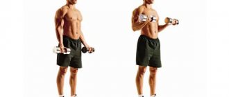
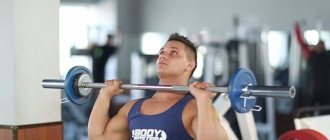
The triceps brachii muscle usually responds well to special exercises. But for some people it is classified as a “lagging behind.” Then it is necessary to pay special attention to the triceps muscle, “punching” it purposefully, in isolation.
Although trainers advise starting classes with basic (multi-joint exercises), if the triceps are lagging, you can “start” differently. There are many techniques for this: pre-fatigue of the muscle, isolation training and others. Only an experienced instructor will tell you how to act in each specific case.
The triceps brachii muscle is often involved when working the pectoral muscles, so this is something to keep in mind. But it is recommended to load no more than twice a week to avoid overtraining of muscle fibers.
The exercises themselves can be easily found in bodybuilding manuals or on websites on the Internet. Many training complexes are posted online on video channels.
Recommendations for brachialis training
Since the increase in biceps directly depends on the growth of the brachialis, which pushes out the external muscle, arm training must necessarily be accompanied by exercises for the flexors with different grips, and most importantly - neutral and with different equipment.
Thus, not only the usual lifting of barbells and dumbbells should constitute a training plan. A forgotten and rarely used piece of equipment, but very effective for working the shoulder muscles, is the kettlebell. By grasping the core or arms of the kettlebell with a parallel grip, lifting the weight is done more with the help of the brachialis, rather than the biceps. You can also use a barbell for a neutral grip, and even plates, which also provide the necessary grip and load.
You should not train your biceps and brachialis muscles on a separate day more than once a week. Don't forget about the existence of the torso and leg muscles. The fact is that the brachialis is involved in exercises for the back, including pull-ups and rows of barbells and dumbbells to the belt. During leg exercises such as deadlifts or deadlifts, the brachialis also experiences static load when holding weight. Therefore, the muscle must have time to recover; as a synergist, the shoulder muscle experiences a large and regular load while strengthening many muscles of the body. Three exercises for the biceps and brachialis are enough, in which 3-4 sets of 8 to 12 repetitions are performed until failure.
About muscle pain
Unfortunately, the triceps brachii muscle can experience unpleasant sensations, the functions of which lead to overstrain and spasm. Pain may occur when pushing heavy objects or sharply straightening the arm.
Trigger points and muscle bands can also cause problems. In the first case, the pain can radiate to other areas, in the second, it will be very painful on palpation.
In such cases, stretching of the heads of the triceps muscle is required. A massage therapist can do a great job with this.
Read also[edit | edit code]
- Muscles - anatomy and functions
- Arm muscles
- Triceps - exercises and training features
- Triceps brachii muscle - training and exercises
- Biceps brachii
- Brachialis muscle
- Brachioradialis muscle
- Elbow muscle
- Arch support
- Pronator quadratus
- Pronator teres
- Extensor carpi radialis
- Extensor carpi ulnaris
- Flexor carpi radialis and palmaris longus muscles
- Flexor carpi ulnaris
Shoulder joint:
The shoulder joint has the greatest mobility in our body. With it we can rotate our arms in different positions. Agree that it is freedom of movement that provides us with a sense of the fullness of life.
In the shoulder joint, a special classification of tissues can be distinguished, which are referred to as “soft”. These tissues are responsible for joint mobility and also stabilize the joint. Soft tissues are very vulnerable and are often subject to wear and tear, causing injury to the shoulder joint.
Soft tissues include:
- Joint capsule
- Shoulder ligaments
- Superior labrum
- Long head of biceps tendon
- Rotator cuff
- Bursa
The head of the humerus performs a very important function - it is responsible for maintaining the stability of the entire joint, and it is located in the very center of the articular capsule. The humerus is held in position by ligaments, tendons, and anterior muscles.