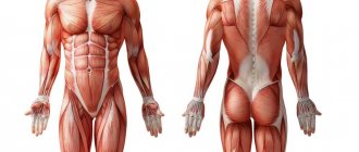
The muscular system is the foundation of physical health. The anatomy of human muscles is represented by more than 600 different fibers, which make up up to 47% of the total mass of the body. Not only the movement of the body in space, but also many physiological processes depend on their functionality: swallowing, blood circulation, chewing, metabolism, heart contractions, etc. The muscular frame forms the structure of the body, ensures position relative to surrounding objects, and allows a person to take part in various physical activities and perform most of the work. Therefore, a detailed study of the structure of muscles, their classification and functionality is considered one of the key sections of anatomy.
Detailed structure of muscle tissue
Each individual muscle is an integral organ consisting of many small muscle fibers - myocytes, as well as dense and loose connective tissue in varying proportions. It has 2 functional zones: the abdomen and the tendon. The abdomen performs mainly a contractile function, therefore it is represented by a combination of connective tissue and myocytes capable of contraction and excitation. The tendon is considered the passive part of the muscle. It is located along the edges and consists of dense connective tissue, thanks to which fibers are attached to bones and joints.
Innervation and blood supply to each muscle is carried out due to the thinnest capillaries and nerve fibers located between bundles of 10–50 myocytes. Thanks to this, muscle tissue receives the necessary nutrition, is supplied with oxygen and nutrients, and can also contract in response to an impulse transmitted by nervous tissue.
Each muscle fiber looks like a long multinucleated cell, the length of which is several times greater than the cross section. The membrane covering the myocyte combines a different number of small myofibrils, depending on the number of which, white and red muscles are distinguished. In white myocytes, the number of myofibrils is higher, so they respond faster to impulses and contract more actively. Red fibers belong to the group of slow fibers because they have fewer myofibrils.
Each myofibril consists of a number of substances on which the functional characteristics and properties of muscles depend:
- Actin is an amino acid protein structure capable of contraction.
- Myosin is the main component of myofibrils, formed by polypeptide chains of amino acids.
- Actinomyosin is a complex of protein molecules actin and myosin.
The main part of myocytes consists of proteins, water and auxiliary components: salts, glycogen, etc. Moreover, the majority is water - its percentage ranges from 70–80%. Despite this, each individual muscle fiber is extremely strong and resilient, and this strength increases depending on the number of myocytes combined into the muscle.
What does strength depend on, besides muscle size?
The larger the muscle, the thicker its fibers and the more force it can produce during contraction. This is why bodybuilders are stronger than untrained people. But at the same time, they are weaker than strength athletes, who have the same or less muscle mass. This means that, in addition to the volume of muscle fibers, there are other factors that influence force production.
Function of the nervous system
For a muscle to begin contracting, the brain must give a signal. An electrical impulse will leave the motor cortex, reach the spinal cord, and from there along the fibers of the motor neurons it will reach the muscle and force its fibers to work.
The more fibers in a muscle contract, the more force a person can produce. Most untrained people cannot voluntarily tense 100% of the fibers. Even with the greatest effort, only about 90% will work.
Strength training increases the nervous system's ability to fire more muscle fibers. In this case, only really heavy loads are used - with 80% of the maximum possible weight. The Why strength depends on more than muscle study found that three weeks of training at 80% of one-repetition maximum (1RM) increased muscle fiber recruitment by 2.35%, while training with light weights - 30% of 1RM - had a negligible effect - just 0.15%.
Moreover, exercise with heavy weights generally increases muscle efficiency.
Tendon stiffness
When a muscle contracts, energy is transferred to a tendon, a dense connective tissue that attaches muscles to bones and moves joints. If the tendon is very stiff, it will prevent the muscle from becoming shorter before the joint angle changes. In this case, muscle contraction and movement in the joint occur simultaneously.
If the tendon is not rigid, during contraction the muscle shortens faster than the bend angle changes. The tendon lengthens and allows the muscle to become shorter before the limb bends at the joint. This increases the speed of contraction but decreases the force.
Strength training increases Human tendon adaptation in response to mechanical loading: a systematic review and meta-analysis of exercise intervention studies on healthy adults tendon stiffness, and working with heavy weights - up to 90% of the one-repetition maximum - gives better results.
Ability to activate the right muscles
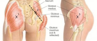
All muscles in our body are interconnected. For example, the biceps is involved in flexing the shoulder joint, and the triceps is involved in extending it. The rectus muscle is responsible for flexion of the hip joint, and the gluteal muscles are responsible for extension. Muscles with such opposite effects are called antagonists.
In order for the force to be maximum during movement, the working muscles (agonists) must tense, and the opposite ones (antagonists) must relax, otherwise they will interfere. Repeating the same movements over and over again improves coordination and the ability to tense and relax the right muscles.
Therefore, strength training is quite monotonous: athletes improve their skills in one movement and perform it better and better.
Bodybuilders, on the contrary, often change exercises, joint flexion angles and machines so that the muscles do not get used to it, and the body constantly experiences the stress necessary for their growth.
In addition, during complex multi-joint movements, in addition to agonists, other muscles are activated - synergists, which increase stability and help produce more force. For example, during squats, the main work is done by the leg muscles, but the abs are also involved. Without his strong muscles, the results in the squat will be much more modest.
Therefore, to be strong, you need to work all the muscles of the body involved in a particular movement. For example, bodybuilders who work only for mass often have fairly developed chests, shoulders and arms, but they pay less attention to the core muscles. Strength athletes, on the contrary, have developed back extensor muscles, core muscles, and buttocks - they increase body stability and help develop more strength during movements.
Muscle anatomy: classification and functions
A huge number of muscles in anatomy are classified according to various criteria, including structure, physiological characteristics, shape, size, location and other indicators. Let's look at each group to understand how human muscle tissue is structured:
- Smooth muscle fibers are a structural unit of the walls of internal organs, blood capillaries and vessels. They contract and relax regardless of the impulses sent by the human consciousness. The work of smooth muscles is distinguished by consistency, regularity and continuity.
- Skeletal muscles are the framework of the human body. They are responsible for physical activity, maintaining the body in a certain position and motor capabilities of a person. The activity of skeletal muscles is controlled by the brain. Myocytes of this group quickly contract and relax, actively respond to training, but are prone to fatigue.
- Cardiac muscle is a separate type of myocyte that combines some of the functional features of smooth and skeletal fibers. On the one hand, its activity is continuous and does not depend on nerve impulses sent by consciousness, and on the other, contractions are carried out quickly and intensely.
Also, muscles are divided into topographic groups based on their location. The body contains muscles of the lower extremities (foot, thigh and lower leg), upper extremities (hand, shoulder and forearm), as well as the head, neck, chest, back and abdomen. Each of these groups is divided into deep and superficial, external and internal.
Depending on the number of joints covered by the muscle, they are divided into single-joint, double-joint and multi-joint. The more joints involved, the higher the functionality of a particular muscle.
In addition, muscles are classified by shape and structure. The simple group includes spindle-shaped, long, straight, short and wide fibers. Multicipital muscles are complex. They are represented by the biceps, consisting of 2 heads, triceps - of 3 heads and quadriceps - of 4 heads. In addition, multitendon and digastric groups of myocytes are considered complex. They are square, deltoid, pyramidal, jagged, diamond-shaped, soleus-shaped, round or triangular.
Depending on the functional features, the following are distinguished:
- flexors,
- extensors,
- pronators (rotators in the medial direction),
- instep supports (rotators to the outside),
- muscles responsible for abduction and adduction, raising and lowering, etc.
The bulk of the muscles work in pairs, performing a common or opposite function. The agonist muscle performs a certain action (for example, flexion), and the antagonist does the exact opposite (that is, extension). Such a complex multi-stage complex ensures coordinated and smooth movements of the human body.
Reduction modes. Strength and muscle function
The following modes of muscle contraction are distinguished:
1. Isotonic contractions. The length of the muscle decreases, but the tone does not change. They do not participate in the motor functions of the body.
2. Isometric contraction. The length of the muscle does not change, but the tone increases. They form the basis of static work, for example when maintaining body posture.
3. Auxotonic contractions. Both the length and tone of the muscle change. With their help, body movement and other motor acts occur.
Maximum muscle strength
- this is the maximum tension that a muscle can develop. It depends on the structure of the muscle, its functional state, initial length, as well as gender, age, and level of training of the person.
Depending on the structure, muscles with parallel fibers (for example, sartorius), fusiform (biceps brachii), and feathery (gastrocnemius) are distinguished. These muscle types have different physiological cross-sectional areas
is the sum of the cross-sectional areas of all the muscle fibers that form the muscle. The largest physiological cross-sectional area, and, consequently, strength, is found in the pennate muscles. The smallest in muscles with parallel fibers.
With moderate stretching of the muscle, the force of its contraction increases, but with overstretching it decreases. With moderate heating, the strength also increases, and with cooling it decreases. Muscle strength decreases due to fatigue, metabolic disorders, etc. The maximum strength of various muscle groups is determined by dynamometers (wrist, deadlift, etc.).
To compare the strength of different muscles, their specific or absolute strength
. It is equal to the maximum force divided by square meters. see muscle cross-sectional area. The specific strength of the human gastrocnemius muscle is 62 kg/cm2, the triceps muscle is 16.8 kg/cm2, and the masseter muscle is 10 kg/cm2.
Muscle work is divided into dynamic and static Dynamic
performed when moving cargo.
During dynamic work, the length of the muscle and its tension change. Therefore, the muscle works in an auxotonic mode. During static
operation, the load does not move, i.e. the muscle works in isometric mode.
Dynamic work is equal to the product of the weight of the load and the height of its lifting or the amount of muscle shortening (A = M h). Work is measured in kg m, joules. The dependence of the amount of work on the load obeys the law of average loads. As the load increases, muscle work initially increases. At medium loads it becomes maximum. If the increase in load continues, the work decreases. The size of the work is also influenced by its rhythm. Maximum muscle work is carried out at an average rhythm. Of particular importance in calculating the amount of workload is the definition of muscle power - this is the work performed per unit of time (P = A·T). The unit of measurement is watt (W).
Muscle fatigue
Fatigue is a temporary decrease in muscle performance as a result of work. Fatigue of an isolated muscle can be caused by its rhythmic stimulation. As a result, the strength of contractions progressively decreases. The higher the frequency, strength of irritation and magnitude of the load, the faster fatigue develops. With fatigue, the single contraction curve changes significantly. The duration of the latent period, the shortening period and especially the relaxation period increases, but the amplitude decreases. The stronger the muscle fatigue, the longer the duration of these periods. In some cases, complete relaxation does not occur. Contracture develops
is a state of prolonged, involuntary muscle contraction.
Muscle work and fatigue are studied using ergography. In the last century, based on experiments with isolated muscles, 3 theories of muscle fatigue were proposed.
1. Schiff's theory: fatigue is a consequence of depletion of energy reserves in the muscle.
2. Pflueger’s theory: fatigue is caused by the accumulation of metabolic products in the muscle.
3. Verworn's theory: fatigue is explained by a lack of oxygen in the muscle.
Indeed, these factors contribute to fatigue in experiments on isolated muscles. ATP resynthesis is disrupted in them, lactic and pyruvic acids accumulate, and the oxygen content is insufficient. However, in the body, intensively working muscles receive the necessary oxygen, nutrients, and are freed from metabolites due to increased general and regional blood circulation. Therefore, other theories of fatigue have been proposed. neuromuscular junctions play a role in fatigue.
.
Fatigue at the synapse develops due to depletion of neurotransmitter stores. However, the main role in fatigue of the musculoskeletal system belongs to the motor centers of the central nervous system. In the last century I.M. Sechenov found that if the muscles of one arm become fatigued, their performance is restored faster when working with the other arm or legs. He believed that this was due to the switching of excitation processes from one motor center to another. He called rest with the inclusion of other muscle groups active
.
It has now been established that motor fatigue is associated with inhibition of the corresponding nerve centers, as a result of metabolic processes in neurons, deterioration in the synthesis of neurotransmitters, and inhibition of synaptic transmission.
Motor units
The main morpho-functional element of the neuromuscular system of skeletal muscles is the motor unit (MU). It includes the spinal cord motor neuron with the muscle fibers innervated by its axon. Inside the muscle, this axon forms several terminal branches. Each such branch forms a contact - a neuromuscular synapse on a separate muscle fiber. Nerve impulses coming from a motor neuron cause contractions of a specific group of muscle fibers. Motor units of small muscles that perform fine movements (muscles of the eye, hand) contain a small number of muscle fibers. In large ones there are hundreds of times more of them.
All MUs, depending on their functional characteristics, are divided into 3 groups:
I. Slow and tireless. They are formed by “red” muscle fibers, which have fewer myofibrils. The contraction speed and strength of these fibers are relatively small, but they are not easily fatigued. Therefore, they are classified as tonic. The regulation of contractions of such fibers is carried out by a small number of motor neurons, the axons of which have few terminal branches. An example is the soleus muscle.
II B. Fast, easily tired. Muscle fibers contain many myofibrils and are called “white.” They contract quickly and develop great strength, but tire quickly. That's why they are called phase
. The motor neurons of these motor units are the largest and have a thick axon with numerous terminal branches. They generate high frequency nerve impulses. For example, the muscles of the eye.
II A. Fast, resistant to fatigue. They occupy an intermediate position.
Smooth muscle physiology
Smooth muscles are present in the walls of most digestive organs, blood vessels, excretory ducts of various glands, and the urinary system. They are involuntary and provide peristalsis of the digestive and urinary systems, maintaining vascular tone.
Unlike skeletal muscles, smooth muscles are formed by cells that are often spindle-shaped and small in size, without transverse striations. The latter is due to the fact that the contractile apparatus does not have an ordered structure. Myofibrils consist of thin filaments of actin that run in different directions and attach to different parts of the sarcolemma. Myosin protofibrils are located next to actin ones. The elements of the sarcoplasmic reticulum do not form a system of tubes. Individual muscle cells are connected to each other by contacts with low electrical resistance - nexuses
, which ensures the spread of excitation throughout the smooth muscle structure. The excitability and conductivity of smooth muscles is lower than that of skeletal muscles.
The membrane potential is 40-60 mV, since the membrane of smooth muscle cells (SMCs) has a relatively high permeability to sodium ions. Moreover, in many smooth muscles the membrane potential (MP) is not constant. It periodically decreases and returns to its original level. Such oscillations are called slow waves
(MV).
When the peak of the slow wave reaches a critical level of depolarization, action potentials (APs) begin to be generated on it, accompanied by contractions. MV and AP are carried out through smooth muscles at a speed of only 5-50 cm/sec. Such smooth muscles are called spontaneously active
, because. they are automatic. For example, due to such activity, intestinal peristalsis occurs. The pacemakers of intestinal peristalsis are located in the initial sections of the corresponding intestines.
The generation of AP in SMCs is due to the entry of calcium ions into them. The electromechanical coupling mechanisms are also different. Contraction develops due to calcium entering the cell during AP. calmodulin, mediates the connection between calcium and myofibril shortening.
.
The contraction curve is also different. The latent period, the period of shortening, and especially relaxation, is much longer than that of skeletal muscles. The contraction lasts several seconds. Smooth muscles, unlike skeletal muscles, are characterized by the phenomenon of plastic tone
– this ability is in a state of contraction for a long time without significant energy consumption and fatigue.
Thanks to this property, the shape of internal organs and vascular tone are maintained. In addition, smooth muscle cells themselves are stretch receptors. When they are tensioned, PDs begin to be generated, which leads to contraction of the SMC. This phenomenon is called the myogenic mechanism of regulation of contractile activity
.
Physiology of human muscles
The main properties of muscle tissue that ensure full functionality of the structures include:
- Contractility is the ability to contract.
- Excitability is a reaction to a nerve impulse.
- Elasticity is a change in the length and diameter of fibers depending on external and internal influences.
Muscle contraction is regulated through the activity of the nervous system. Each muscle contains many nerve endings, which can be divided into 2 types - receptors and affectors. Sensitive receptors perceive the speed and degree of stretching and contraction, the force of impact and movement of myocytes. They can be located freely, branching in the thickness of the muscle, or not freely, intertwining into a fusiform complex. Information about the state and position of the muscle fiber from the receptors enters the central nervous system, from where it is transmitted back to the effectors, causing their excitation and, as a result, a reaction to the received impulse.
Myocyte contraction occurs due to the penetration of actin filaments between myosin chains. In this case, the total length of actin and myosin fibers does not change - the contraction occurs due to a change in the length of the actinomyosin complex. This mechanism is called sliding and is accompanied by the consumption of the body’s energy reserves.
Muscles also contain nerve fibers that regulate the metabolic process and the state of myocytes at rest. Thanks to this, the functioning of muscle tissue is regulated, overwork and unphysiological overstretching or contraction are prevented. This mechanism allows you to adapt muscle work to the environment and ensure full functionality of the body.
Conclusion
The anatomy of muscles, their number and ratio is physiologically unchanged, depending on heredity and characteristics of the body. However, with the right amount of exercise, regular exercise and a healthy lifestyle, you can develop muscle fibers, greater endurance, strength and resilience. You should not assume that only the condition of the skeletal muscles and the relief of the body depend on this - a properly composed set of exercises also improves the functioning of smooth and cardiac myocytes. Thanks to this, you can start a “feedback” cycle: the heart muscle, developed through regular training, better pumps blood throughout the body, so all organs, including skeletal muscles, receive more nutrition and oxygen necessary to overcome stress. And physically developed skeletal and smooth muscles, in turn, better support internal organs, ensuring their full functioning.
Knowing the basics of human muscle anatomy, you can competently build a training process, introduce the basics of physical activity into your life and at the same time improve the condition of the body as a whole.